Neurofilament light and glial fibrillary acidic protein do not reflect neuronal or glial damage during different intracranial radiotherapy regimes: a pilot study - Report - MDSpire
Advertisement
Neurofilament light and glial fibrillary acidic protein do not reflect neuronal or glial damage during different intracranial radiotherapy regimes: a pilot study
Clinical Report: Neurofilament Light Chain and GFAP Levels in Radiotherapy
Overview
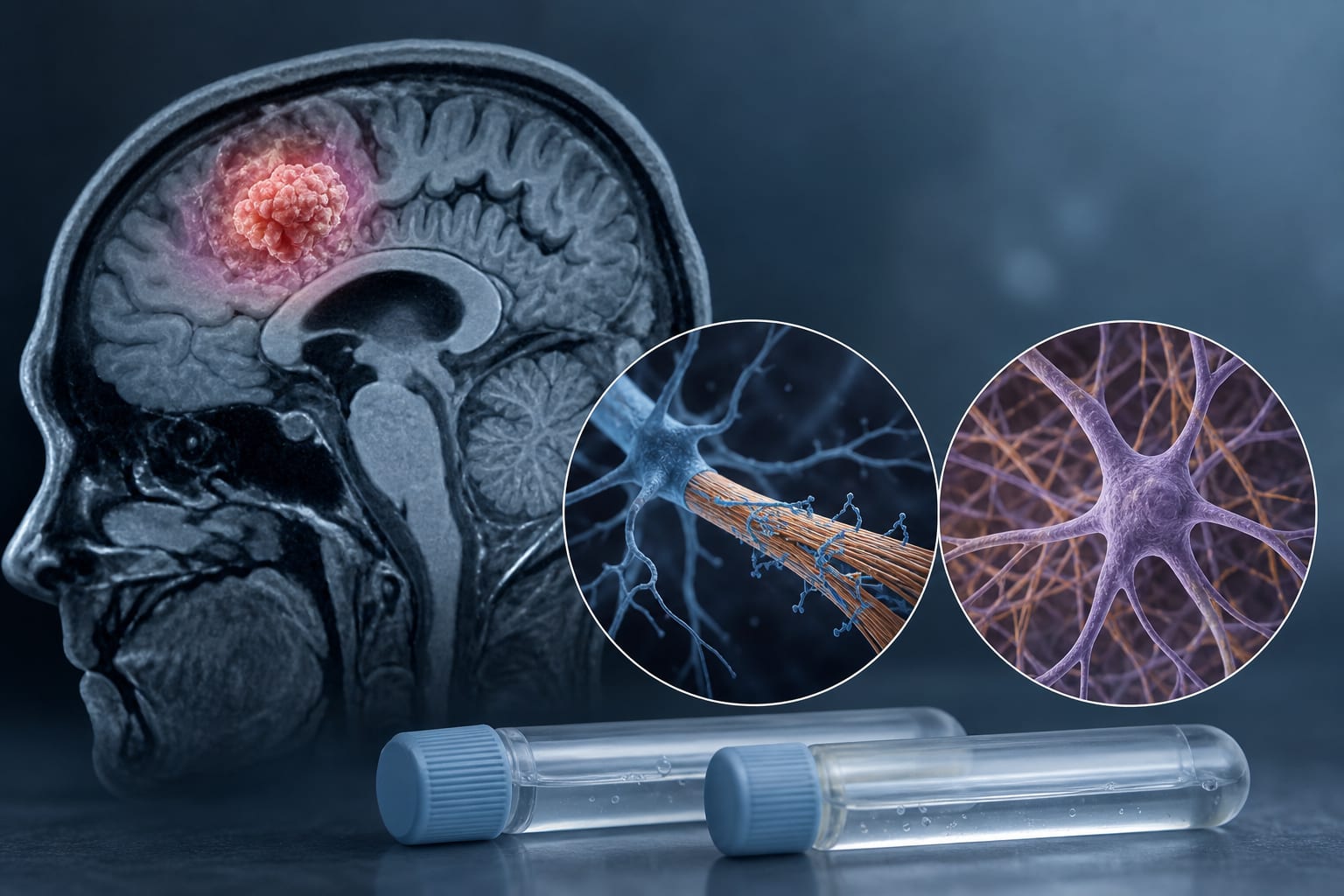
This pilot study investigates the levels of neurofilament light chain (NfL) and glial fibrillary acidic protein (GFAP) in patients undergoing various intracranial radiotherapy protocols. The findings indicate that while NfL and GFAP levels are elevated prior to treatment, significant increases during radiotherapy are not observed, suggesting limited utility as markers for cerebral radiation injury.
Background
Neurofilament light chain and glial fibrillary acidic protein are established non-specific markers of central nervous system damage, often associated with brain tumors. Understanding their response to radiotherapy is crucial, as it may help in monitoring treatment effects and detecting new lesions in patients with cerebral metastases.
Data Highlights
No significant numerical data or trial results were presented in the source material.
Key Findings
Elevated NfL and GFAP levels were confirmed in all patients before the start of radiotherapy.
No significant increase in NfL and GFAP levels was observed after the onset of radiotherapy.
Long-term follow-up showed that decreasing NfL and GFAP values correlated with treatment response.
Pronounced increases in serum NfL levels were associated with the detection of new cerebral lesions.
The study suggests that NfL and GFAP may be suitable for follow-up and early detection of new lesions post-treatment.
Clinical Implications
The findings suggest that NfL and GFAP may not be reliable indicators of neuronal or glial injury during radiotherapy. However, their potential utility in monitoring treatment response and detecting new lesions warrants further investigation.
Conclusion
In summary, NfL and GFAP levels do not correlate with cerebral radiation injury during treatment, but they may serve as useful markers for follow-up assessments.
by Yvonne Dzierma, Holger Sebb, Michael Utzig, Nurlan Abdullayev, Christian Berdel, Christian Ruebe, Jochen Fleckenstein, Markus Hecht, Guido Hildebrandt, Mathias Jucker, Kristina Heyne