Habitat-Radiomics combining multichannel 2.5D deep learning for differentiating adrenal adenomas from metastases using automatic segmentation: a multicenter study - Report - MDSpire

Habitat-Radiomics combining multichannel 2.5D deep learning for differentiating adrenal adenomas from metastases using automatic segmentation: a multicenter study

  • By

  • shengnan, Yin

  • Ning, Ding

  • Chuqi, Yang

  • Shaocai, Wang

  • Mengjuan, Li

  • Ji, Yiding

  • Tong, Liu

  • Long, Jin

  • May 27, 2026

  • 0 min

Share

Clinical Report: Integrating Habitat-Radiomics with Multichannel 2.5D Deep Learning

Overview

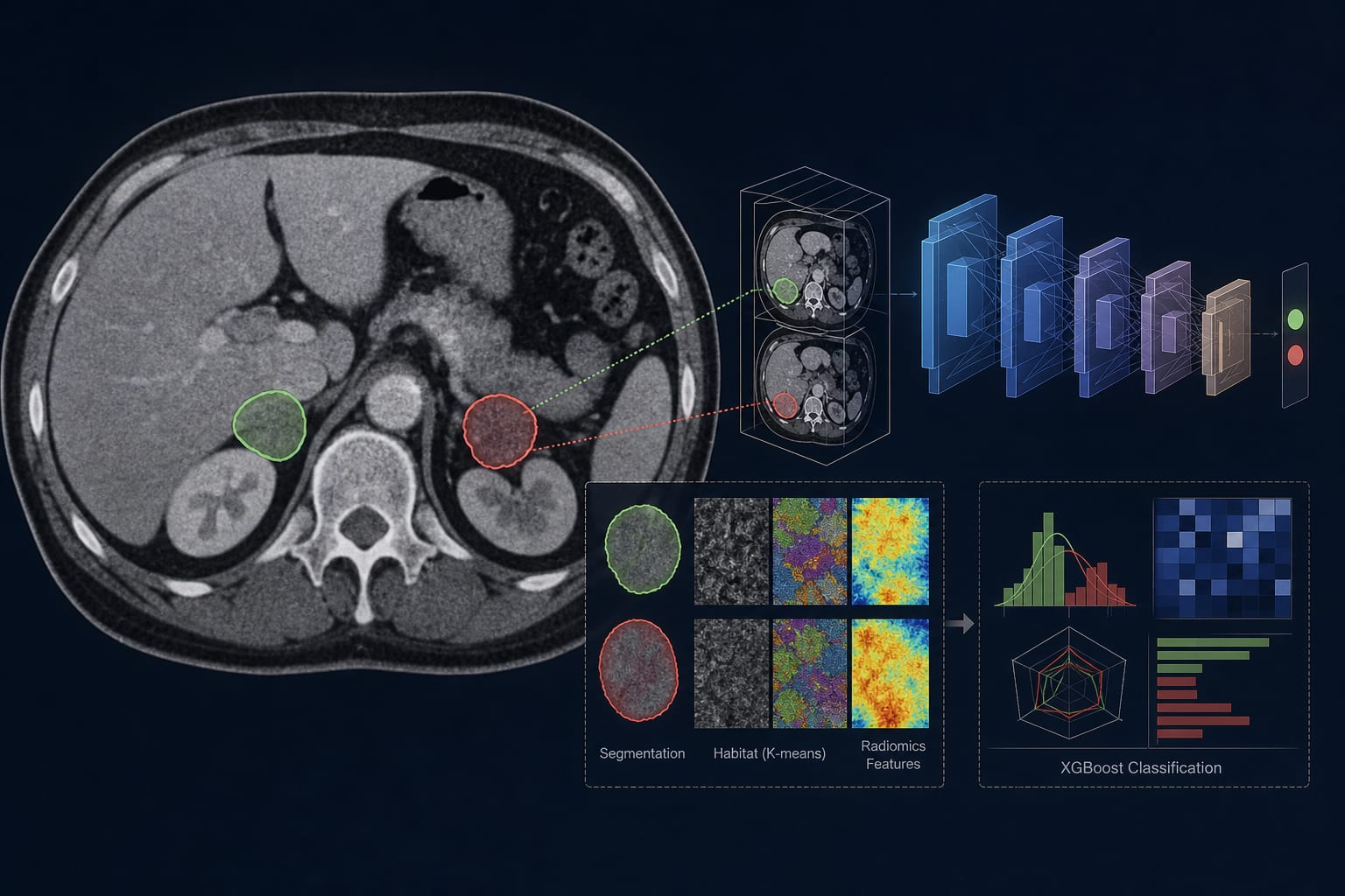
This study evaluates a fusion model that integrates habitat-radiomics and deep learning for the automated differentiation of lipid-poor adrenal adenomas from metastatic lesions. The model demonstrated high predictive performance, with AUCs reaching 0.983 in the training set.

Background

Differentiating between lipid-poor adrenal adenomas and metastatic lesions is clinically significant due to differing treatment approaches and prognoses. The increasing prevalence of adrenal incidentalomas necessitates improved diagnostic tools to guide management decisions. Advances in imaging and machine learning techniques, such as radiomics and deep learning, offer potential solutions to these diagnostic challenges.

Data Highlights

ModelAUC (Training)AUC (Internal Validation)AUC (External Test)
Fusion Model0.9830.9130.886
2.5D Deep Learning0.830–0.945--
Habitat-Radiomics0.825–0.970--

Key Findings

  • The fusion model achieved an AUC of 0.983 in the training set.
  • Standalone models showed AUCs ranging from 0.830 to 0.945 for the 2.5D deep learning model.
  • Habitat-radiomics features contributed to the predictive performance with AUCs from 0.825 to 0.970.
  • The study involved a total of 390 patients across two hospitals.
  • Automated segmentation was performed using the Medical SAM model.

Clinical Implications

The fusion model may assist clinicians in noninvasively differentiating between adrenal adenomas and metastatic lesions, potentially guiding treatment decisions. The integration of advanced imaging techniques and machine learning could enhance diagnostic accuracy in adrenal pathology.

Conclusion

The findings suggest that the fusion model has significant potential for improving the differentiation of adrenal lesions, which is crucial for appropriate clinical management.

Related Resources & Content

  1. European Radiology, 2023 -- Utilizing Radiomics for Enhanced Characterization of Lipid-Deficient Adrenal Adenomas in Unenhanced CT Imaging
  2. European Radiology, 2024 -- Evaluating the Diagnostic Efficacy of Photon-Counting Detector CT in Distinguishing Adrenal Adenomas from Metastatic Lesions
  3. European Radiology, 2023 -- Automated MRI Segmentation and Radiomic Feature Analysis of Hypopharyngeal Cancer Utilizing Deep Learning Techniques
  4. European Journal of Endocrinology, 2023 -- European Society of Endocrinology clinical practice guidelines on the management of adrenal incidentalomas
  5. ScienceDirect, 2017 -- Management of Incidental Adrenal Masses: A White Paper of the ACR Incidental Findings Committee
  6. Enhancing Lesion Evaluation in Longitudinal CT Imaging: A Multi-Center Study on AI-Enhanced Registration and Volumetric Segmentation Techniques
  7. ACR Incidental Findings
  8. Artificial intelligence and radiomics applications in adrenal lesions: a systematic review
  9. European Society of Endocrinology clinical practice guidelines on the management of adrenal incidentalomas, in collaboration with the European Network for the Study of Adrenal Tumors | European Journal of Endocrinology | Oxford Academic
  10. Management of Incidental Adrenal Masses: A White Paper of the ACR Incidental Findings Committee - ScienceDirect
  11. Adrenal Neoplasms: Lessons from Adrenal Multidisciplinary Tumor Boards | RadioGraphics
  12. Transformer-based integration of radiomics and deep learning for differentiating lipid-poor adrenal adenomas from malignant tumors - ScienceDirect
  13. Frontiers | Multi-model radiomics and machine learning for differentiating lipid-poor adrenal adenomas from metastases using automatic segmentation
  14. Evaluation of a fusion model combining deep learning models based on enhanced CT images with radiological and clinical features in distinguishing lipid-poor adrenal adenoma from metastatic lesions | BMC Medical Imaging | Full Text
  15. Deep Learning for Adrenal Gland Segmentation: Comparing Accuracy and Efficiency Across Three Convolutional Neural Network Models | MDPI
  16. A deep learning algorithm for automated adrenal gland segmentation on non-contrast CT images | BMC Medical Imaging | Springer Nature Link

Original Source(s)

Related Content