Clinical Report: Paucibacillary Tuberculous Meningitis and Diagnostic Challenges
Background
Tuberculous meningitis (TBM) is the most severe form of tuberculosis, with mortality exceeding 20% even with appropriate treatment. Diagnosis can be particularly challenging when patients lack classical features such as fever, meningeal signs, or pulmonary involvement and when microbiological tests are negative, as may occur in paucibacillary TBM.
Data Highlights
The patient was a 63-year-old woman with newly diagnosed diabetes who presented with sudden-onset headache, dysarthria, right-sided weakness, and two focal seizures.
Her Marais diagnostic score was 2, below the score of ≥6 required for possible TBM.
Peripheral-blood IGRA was positive at 48.55 pg/mL.
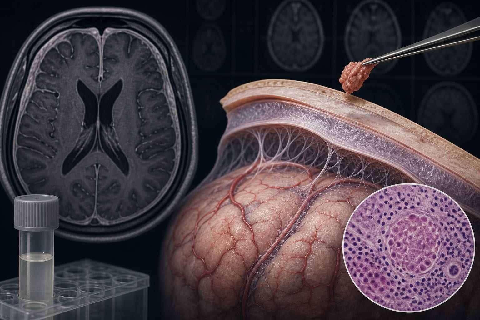
A single acid-fast bacillus was identified after examination of more than 300 high-power fields across three tissue sections.
Anti-tuberculosis therapy lasted approximately 12 months, while follow-up MRI at 9 months showed almost complete resolution of meningeal enhancement and cerebral edema.
Key Findings
The patient lacked typical infectious symptoms and meningeal irritation signs despite having TBM.
CSF GeneXpert MTB/RIF, mycobacterial culture, and metagenomic next-generation sequencing were negative.
Meningeal biopsy demonstrated granulomatous inflammation, and identification of an acid-fast bacillus together with the positive IGRA strongly supported tuberculous etiology.
Normal or near-normal CSF findings do not exclude paucibacillary TBM.
Clinical Implications
TBM should remain a consideration in patients with atypical neurologic presentations even when CSF findings are near normal and molecular or microbiological testing is negative. The authors emphasize that radiological progression despite clinical quiescence should prompt strong consideration of meningeal biopsy, particularly when molecular tests on CSF remain negative.
Conclusion
This case demonstrates that paucibacillary TBM can mimic stroke, viral encephalitis, or noninfectious conditions and may evade contemporary CSF diagnostic testing. Complete symptomatic improvement does not exclude TBM, and tissue biopsy may be important when radiological abnormalities progress despite clinical recovery.
Related Resources & Content
Heemskerk AD, Bang ND, Mai NT, Chau TT, Phu NH, Loc PP, et al. “Intensified antituberculosis therapy in adults with tuberculous meningitis.” New England Journal of Medicine. 2016;374(2):124–134.
Wilkinson RJ, Rohlwink U, Misra UK, van Crevel R, Thwaites GE. “Tuberculous meningitis.” Nature Reviews Neurology. 2017;13(10):581–598.
Leiva-Ordoñez JE, Quintero B. “Diagnosis of tuberculous meningitis: integrating clinical assessment and molecular diagnostics.” Diagnostics. 2026;16(4):552.
Marais S, Thwaites G, Schoeman JF, Trk ME, Marais BJ. “Tuberculous meningitis: a uniform case definition for use in clinical research.” The Lancet Infectious Diseases. 2010;10(11):803–812.
Bahr NC, Nuwagira E, Evans EE, Cresswell FV, Bystrom PV, Byamukama A, et al. “Diagnostic accuracy of Xpert MTB/RIF Ultra for tuberculous meningitis in HIV-infected adults: a prospective cohort study.” The Lancet Infectious Diseases. 2018;18(1):68–75.
Hou J, Liu XJ, He Y, Zhang YA, Wang MS. “Cerebrospinal fluid findings of infant tuberculous meningitis: a scoping review.” Annals of Medicine. 2022;54(1):2517–2521.