Conexiant’s news site is now MDSpire News. Learn more
Advertisement
ORG Detects Retinal Dysfunction Before Structural Changes
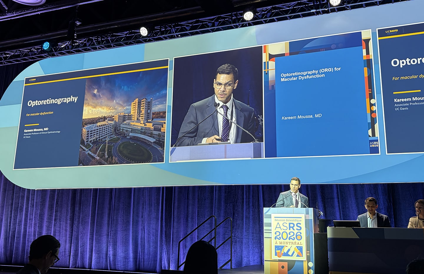
At ASRS 2026, Kareem Moussa, MD, presented early data suggesting optoretinography may identify photoreceptor dysfunction in diabetic eyes before conventional imaging reveals abnormalities.
Clinical Report: ORG Detects Retinal Dysfunction Before Structural Changes
Overview
Optoretinography (ORG) may identify photoreceptor dysfunction in diabetic eyes prior to visible structural changes. Early data presented at the ASRS 2026 meeting indicate that ORG can detect functional abnormalities in eyes that appear structurally normal.
Background
Diabetic retinopathy is a leading cause of blindness, often progressing without early visual symptoms. Current imaging techniques primarily focus on structural changes, which may not reflect the functional status of photoreceptors. The introduction of functional imaging techniques like ORG could enhance early detection and management of retinal diseases.
Data Highlights
Metric
Normal Eyes
Diseased Eyes
V20-40 Elongation Velocity
Decreased with distance from fovea
Attenuated across measured locations
Key Findings
ORG measures photoreceptor responses to light using a standard OCT light source.
Vmin and V20-40 are two velocity-based metrics used to quantify photoreceptor responses.
In diseased eyes, photoreceptor elongation was consistently reduced compared to healthy controls.
ORG detected altered photoreceptor function in structurally normal eyes of diabetic patients.
The technology is still in early development and requires further validation.
Clinical Implications
ORG's ability to detect functional changes in photoreceptors before structural abnormalities may allow for earlier intervention in diabetic retinopathy.
Conclusion
ORG may enable earlier detection of functional impairments in diabetic eyes.
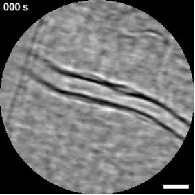
The movement of immune cells through the retinal vasculature has long been inferred from laboratory models. Now, advances in imaging are allowing clinicians to observe these processes directly. At the 2026 meeting of the Association for Research in Vision and Ophthalmology (ARVO) in Denver, Richard B. Rosen, MD, ScD(hon), FACS, FASRS, FARVO, described early clinical work using adaptive optics scanning laser ophthalmoscopy (AOSLO) to visualize leukocyte trafficking in vivo.