Clinical Report: Characterization of Optical Coherence Tomography Findings and Visual Outcomes in Vitreoretinal Lymphoma
Overview
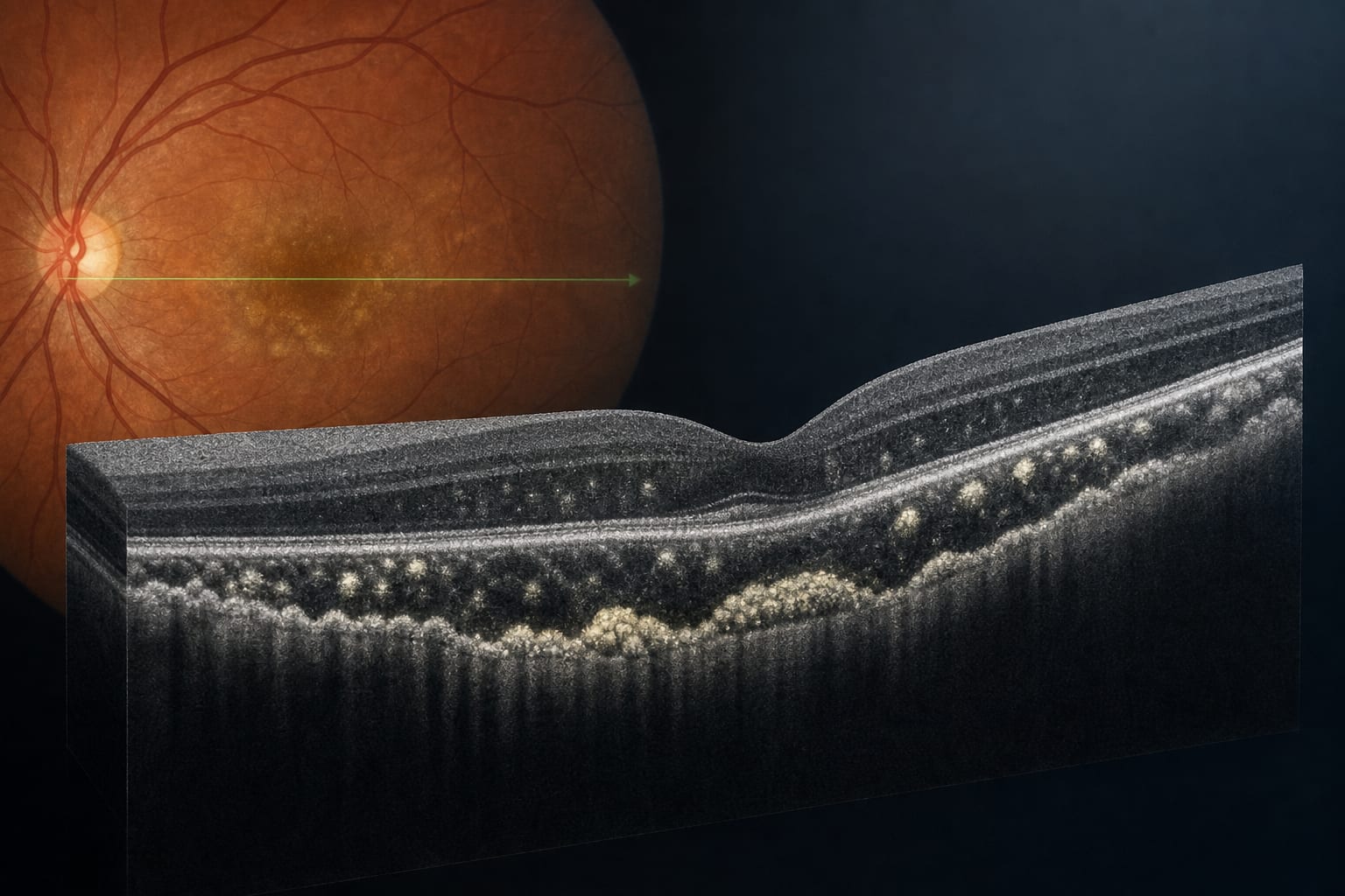
This study characterizes OCT features in vitreoretinal lymphoma (VRL) and identifies associations with visual prognosis. Key findings include the prevalence of sub-RPE deposits and the impact of EZ disruption on visual outcomes following IVT-MTX treatment.
Background
Vitreoretinal lymphoma is an ocular manifestation of primary central nervous system lymphoma, often presenting diagnostic challenges due to its masquerade as inflammatory conditions. The increasing incidence of VRL, particularly in older adults, necessitates effective noninvasive diagnostic tools for early detection and monitoring of treatment response.
Data Highlights
OCT Feature
Prevalence
Sub-RPE deposits
92.1%
Subretinal deposits
65.8%
Intraretinal deposits
23.7%
Key Findings
Sub-RPE deposits were the most prevalent OCT feature in VRL (92.1%).
Baseline best-corrected visual acuity (BCVA) and ellipsoid zone (EZ) disruption were associated with worse visual outcomes.
The LASSO model identified EZ disruption and EZ-RPE attenuation as predictors of significant vision loss.
Following IVT-MTX treatment, vitreous cells resolved rapidly, while sub-RPE and subretinal deposits regressed slowly.
EZ disruption remained unchanged post-treatment, indicating a need for ongoing monitoring.
Clinical Implications
The findings highlight the importance of OCT as a noninvasive tool for assessing VRL and monitoring treatment response. Clinicians should consider baseline OCT features, particularly EZ disruption, when evaluating visual prognosis in VRL patients.
Conclusion
OCT provides valuable insights into the characteristics and treatment responses of VRL, underscoring its role in clinical practice for early detection and ongoing management.