Clinical Report: EGP-Net for Lung Nodule Segmentation Using Advanced Techniques
Overview
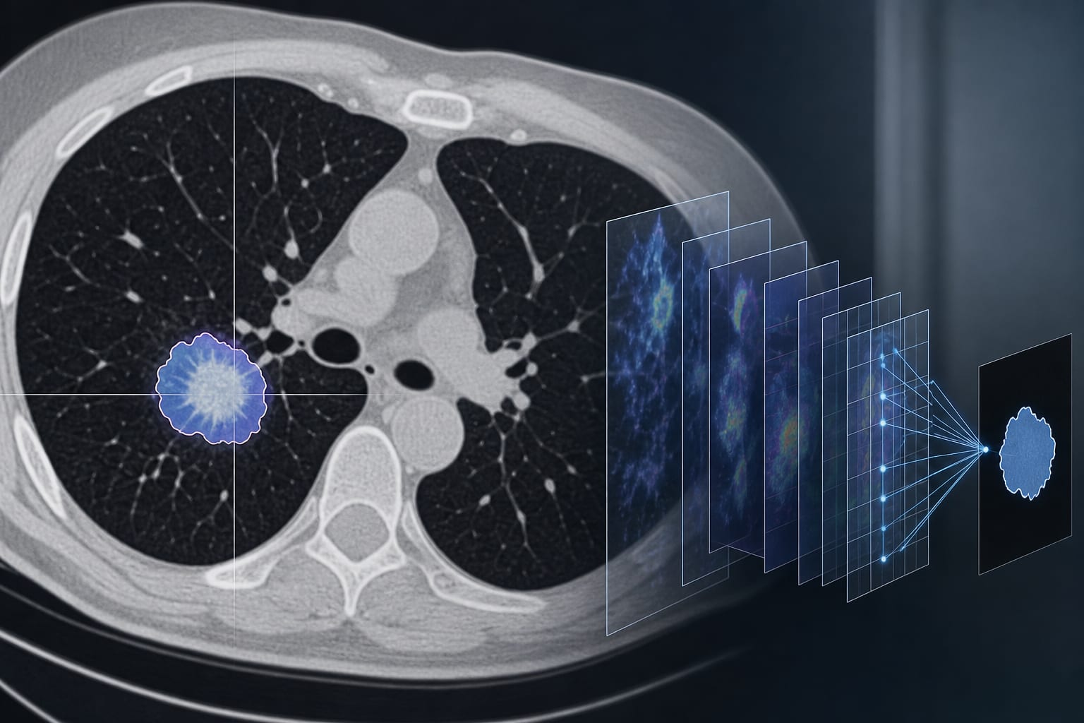
EGP-Net demonstrates significant improvements in the segmentation of pulmonary nodules in CT images, achieving an IoU of 88.32% and a Dice coefficient of 92.65% on the LIDC dataset. The method integrates advanced techniques to enhance accuracy and robustness.
Background
Accurate segmentation of pulmonary nodules is crucial for the early detection and treatment of lung cancer, which remains a leading cause of cancer-related mortality. Traditional manual segmentation methods are time-consuming and subjective, highlighting the need for automated solutions. Recent advancements in deep learning have shown promise in improving segmentation accuracy, yet challenges remain due to variations in nodule characteristics.
Data Highlights
Dataset
IoU
Dice Coefficient
LIDC
88.32%
92.65%
Key Findings
EGP-Net integrates a Res2Net-50 encoder and edge-guided network for enhanced feature representation.
The model utilizes a global pyramid perception module and dynamic attention fusion for improved contextual understanding.
Performance metrics such as IoU, Dice, F2-score, and F0.5-score were used to evaluate segmentation accuracy.
EGP-Net outperformed existing state-of-the-art segmentation methods on the LIDC dataset.
Ablation studies confirmed the effectiveness of each component of the EGP-Net architecture.
Clinical Implications
The EGP-Net model provides an automated approach for the segmentation of pulmonary nodules.
Conclusion
EGP-Net improves the accuracy and robustness of pulmonary nodule segmentation.