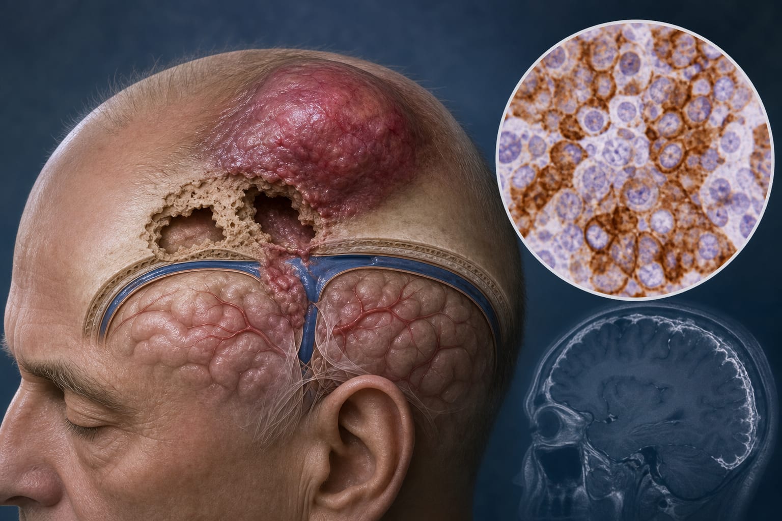
Clinical and Imaging Features of Intracerebral Hemorrhage in Brain Tumors and Metastases
Overview
This study provides a detailed clinical and imaging characterization of intracerebral hemorrhage (ICH) associated with primary brain tumors and metastatic lesions. It highlights the challenges in differentiating neoplastic ICH from other hemorrhagic etiologies and identifies distinct clinical and radiographic features that may aid timely diagnosis and management.
Background
Central nervous system tumors and metastases are among the most lethal cancers, often complicated by neoplastic intracerebral hemorrhage resulting from fragile tumor vessels. Differentiating these hemorrhages from other causes such as hypertensive bleeding is clinically challenging but critical for prompt treatment. Prior studies have explored imaging strategies to improve diagnostic accuracy, yet comprehensive data on incidence and clinical features remain limited. This study aims to fill this gap by analyzing demographics, symptoms, imaging findings, treatment, and outcomes in patients with neoplastic ICH.
Data Highlights
The study retrospectively analyzed patients with confirmed neoplastic intracerebral hemorrhage from a cohort at Charité University Hospital (2016-2020). Clinical data included demographics, Glasgow Coma Scale (GCS), modified Rankin Scale (mRS), vascular risk factors, and prior therapies. Imaging assessments detailed hemorrhage location, volume, perihematomal edema, and presence of intraventricular hemorrhage. Statistical analyses compared tumor-associated ICH and metastasis-associated ICH groups.
Key Findings
Neoplastic ICH patients present with clinical symptoms similar to other cerebrovascular stroke events, including focal deficits, seizures, and altered consciousness.
Distinct clinical and radiographic differences exist between ICH associated with primary brain tumors and metastatic lesions, aiding differential diagnosis.
Imaging revealed that hemorrhages were strictly intratumoral, with variable involvement of cerebral lobes and structures such as basal ganglia and brainstem.
Perihematomal edema and secondary intraventricular hemorrhage were quantified, contributing to understanding hemorrhage severity and prognosis.
Many patients had prior therapies including craniotomy, chemo-, immuno-, or radiotherapy, which may influence hemorrhage characteristics and outcomes.
Modified Rankin Scale dichotomization showed a medically relevant distinction between patients maintaining independence and those requiring complete assistance post-ICH.
Clinical Implications
Clinicians should maintain a high index of suspicion for neoplastic intracerebral hemorrhage in patients with known or suspected brain tumors presenting with acute hemorrhagic stroke symptoms. Detailed imaging evaluation focusing on hemorrhage location and characteristics can facilitate differentiation between tumor-related and non-neoplastic hemorrhages. Early recognition is essential to avoid diagnostic delays and optimize timely therapeutic interventions.
Conclusion
This study enhances understanding of the clinical and imaging profiles of neoplastic intracerebral hemorrhage, emphasizing the importance of distinguishing tumor-related hemorrhages from other causes. Improved diagnostic accuracy may lead to better patient management and outcomes in this high-risk population.
References
Charité Berlin, Germany Study (2016-2020) -- Clinical and Imaging Features of Neoplastic ICH
by Semil Eminovic, Tobias Orth, Andrea Dell’Orco, Lukas Baumgärtner, Andrea Morotti, David Wasilewski, Melisa S. Guelen, Michael Scheel, Tobias Penzkofer, Jawed Nawabi