Epithelial cell proliferation on anterior surface of intraocular lens after Descemet stripping automated endothelial keratoplasty (DSAEK) surgery: a case report - Report - MDSpire
Advertisement
Epithelial cell proliferation on anterior surface of intraocular lens after Descemet stripping automated endothelial keratoplasty (DSAEK) surgery: a case report
Proliferation of Epithelial Cells on the Anterior Surface of Intraocular Lenses
Overview
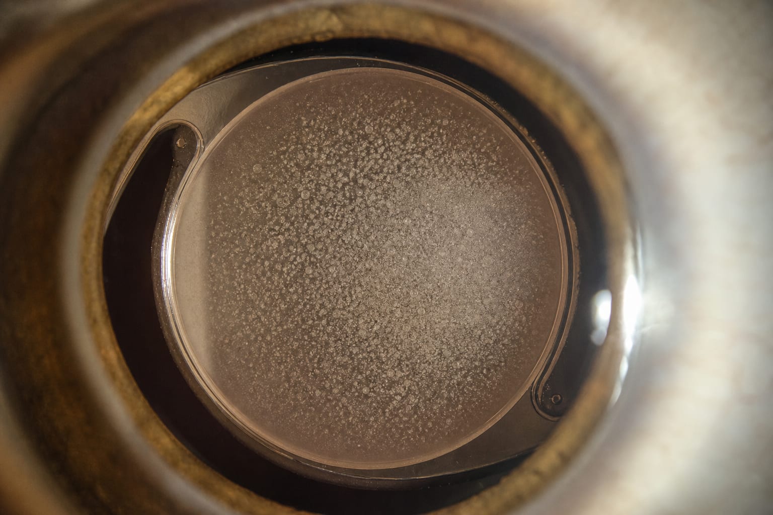
This case study reports a rare instance of epithelial cell proliferation on the anterior surface of an intraocular lens (IOL) following Descemet stripping automated endothelial keratoplasty (DSAEK) in a 61-year-old woman. The patient experienced progressive visual decline due to opacification, which was refractory to initial therapeutic interventions, including Nd: YAG laser therapy.
Background
Intraocular lens (IOL) opacification, though rare, can significantly impair visual function, leading to symptoms such as glare and reduced contrast sensitivity. It can be classified into primary and secondary forms, with secondary opacification often linked to surgical procedures like endothelial keratoplasty. Understanding the mechanisms behind IOL opacification, including the role of residual epithelial cells and environmental factors, is crucial for improving patient outcomes following cataract and corneal surgeries.
Data Highlights
No numerical data or trial data were provided in the article.
Key Findings
Epithelial cell proliferation on IOL surfaces is a rare cause of opacification.
Opacification may arise from residual epithelial cells adhering to the IOL surface post-surgery.
Initial treatments, including Nd: YAG laser therapy, were ineffective in resolving the opacities.
The IOL was ultimately explanted due to firm adhesions with the capsular bag.
Histopathological evaluation of the explanted IOL revealed epithelial-like cellular imprints covering the anterior surface.
Clinical Implications
Clinicians should be aware of the potential for epithelial cell proliferation on IOLs following DSAEK, as it may lead to significant visual impairment. Early recognition and appropriate management strategies are essential to address this complication.
Conclusion
Epithelial cell proliferation on IOLs represents a rare complication following DSAEK, necessitating careful monitoring and intervention.