Clinical Report: Assessment of Retinal Vascular Alterations in DME
Overview
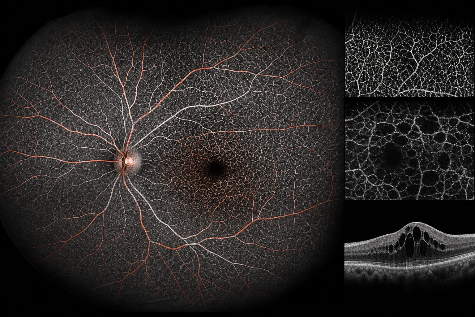
This study evaluates retinal blood flow changes in diabetic macular edema (DME) using ultra-widefield optical coherence tomography angiography (OCTA). Significant differences in vascular density were observed across various diabetic patient groups, highlighting the potential of OCTA in early detection and monitoring of DME.
Background
Diabetic retinopathy (DR) is a leading cause of blindness, making early detection and intervention crucial. Diabetic macular edema (DME) is a significant complication of DR that can severely impair vision. The use of advanced imaging techniques like OCTA allows for detailed assessment of retinal microvasculature, which is essential for managing these conditions effectively.
Data Highlights
Group
Vascular Density Findings
DM
Reduced nasal SVC VD in N11, N16, N21 regions
NPDR
Widespread SVC VD reduction compared to controls and DM
DME
Increased SVC VD in peripheral retina (11–21 mm)
DVC
Significant changes only in macular area for NPDR; increased VD in all regions except I21 for DME
Key Findings
Nasal SVC vascular alterations are detectable early in preclinical DR.
Peripheral DVC VD correlates strongly with the presence of DME.
OCTA provides a non-invasive method for assessing retinal blood flow changes.
Increased peripheral SVC VD in DME patients may indicate altered retinal perfusion.
OCTA can quantitatively analyze retinal features critical for monitoring DME.
Clinical Implications
The findings underscore the importance of using OCTA for early detection of vascular changes in diabetic patients. Clinicians should consider incorporating OCTA into routine assessments to monitor DME progression and guide treatment decisions effectively.
Conclusion
This study highlights the utility of ultra-widefield SS-OCTA in detecting retinal vascular alterations associated with DME. Continued research is needed to further elucidate the relationship between these vascular changes and clinical outcomes in diabetic patients.
Presenting results from the DME AWARE Delphi Study at the Association for Research in Vision and Ophthalmology (ARVO) meeting in Denver, Baruch D. Kuppermann, MD, PhD, director of the Gavin Herbert Eye Institute at the University of California, Irvine, described a set of consensus findings that point to unmet needs across the continuum of DME management, with particular emphasis on early intervention and noninvasive treatment options (Figure 1).