Clinical Report: Approaches to Diagnosing and Managing Airway Complications in Relapsing Polychondritis
Overview
This report reviews the challenges and strategies in diagnosing and managing airway complications associated with relapsing polychondritis (RP). It highlights the importance of early recognition and the role of various diagnostic modalities in improving patient outcomes.
Background
Relapsing polychondritis is a rare autoimmune disease that significantly impacts cartilage, particularly in the airway, leading to severe complications. Airway involvement occurs in approximately 50% of RP patients and is a major cause of morbidity and mortality. Understanding the diagnostic and management strategies for airway complications is crucial for improving patient prognosis.
Data Highlights
No numerical data provided in the source material.
Key Findings
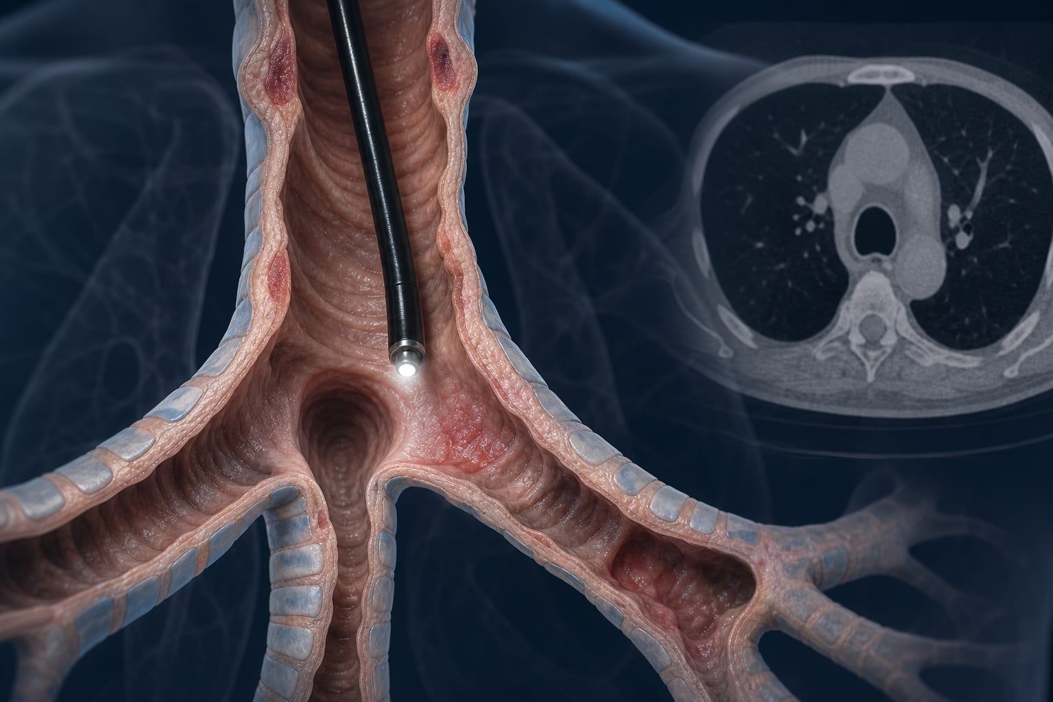
Airway involvement in RP can lead to severe complications such as airway stenosis and tracheomalacia.
Imaging techniques like chest CT and bronchoscopy have high diagnostic value, with positive rates of approximately 88.9% and 85.7%, respectively.
Emerging imaging technologies, including 18F-FDG PET/CT, are beneficial for assessing airway inflammation.
Traditional treatments include glucocorticoids and immunosuppressants, but their efficacy can be limited.
Interventional strategies, such as tracheostomy and airway procedures, are increasingly important in managing severe airway involvement.
Clinical Implications
Clinicians should maintain a high index of suspicion for airway involvement in RP due to its potential severity. A multidisciplinary approach integrating clinical, imaging, and endoscopic evaluations is essential for accurate diagnosis and effective management.
Conclusion
The management of airway complications in relapsing polychondritis requires a comprehensive understanding of the disease and its manifestations. Early diagnosis and a tailored treatment approach can significantly enhance patient outcomes.