Clinical Report: Improved Differentiation of Breast Lesions Using MV-Flow
Overview
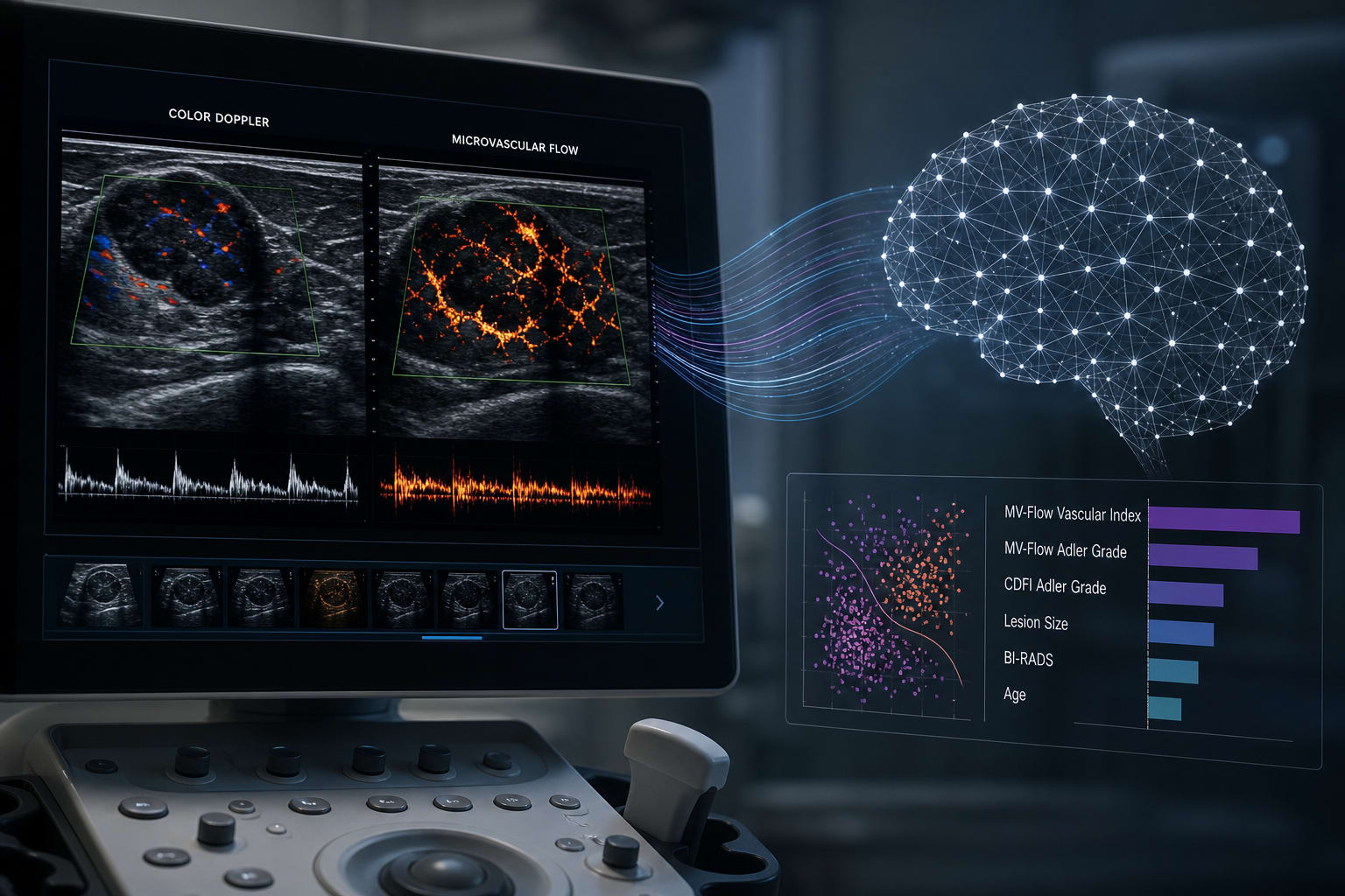
This study demonstrates that Microvascular Flow imaging (MV-Flow) significantly outperforms Color Doppler Flow Imaging (CDFI) in detecting breast lesion microvasculature. Additionally, machine learning models incorporating MV-Flow parameters enhance diagnostic accuracy.
Background
Breast cancer remains a leading cause of morbidity and mortality among women, necessitating effective diagnostic tools for early detection. Conventional imaging techniques, particularly Doppler ultrasound, have limitations in visualizing tumor microvasculature, which is crucial for accurate diagnosis. The integration of advanced imaging modalities like MV-Flow and machine learning may improve the differentiation of breast lesions, thereby impacting clinical decision-making.
Data Highlights
Parameter
MV-Flow
CDFI
Sensitivity
Higher
Lower
Median Vascular Index (VI) in malignant lesions
20.25
N/A
Median Vascular Index (VI) in benign lesions
3.10
N/A
Diagnostic AUC for MV-Flow Adler grade
0.874
N/A
Best ML model accuracy
0.927
N/A
Key Findings
MV-Flow detected blood flow in 16 lesions missed by CDFI.
Inter-observer agreement was significantly higher for MV-Flow (Kappa=0.68) compared to CDFI (Kappa=0.51).
The median Vascular Index (VI) was significantly higher in malignant lesions (20.25) than in benign lesions (3.10, P<0.001).
The combination of MV-Flow Adler grade and VI yielded a diagnostic AUC of 0.888.
The K-Nearest Neighbors model achieved the highest accuracy (0.927) among the eight machine learning models tested.
SHAP analysis identified BI-RADS category and patient age as key predictors in the machine learning model.
Clinical Implications
The findings suggest that MV-Flow could be a valuable tool in clinical practice for improving the accuracy of breast lesion diagnosis. Incorporating machine learning techniques with MV-Flow parameters may further enhance diagnostic capabilities, aiding clinicians in making informed decisions regarding patient management.
Conclusion
MV-Flow represents a significant advancement over traditional CDFI in visualizing breast tumor microvasculature. The integration of machine learning with MV-Flow parameters holds promise for optimizing diagnostic accuracy in breast cancer evaluation.