Longitudinal spatial changes of the contralesional corticospinal tract in high-grade glioma: a within-patient DTI tractography study - Report - MDSpire
Clinical Report: Longitudinal Contralesional CST Changes in High-Grade Glioma
Overview
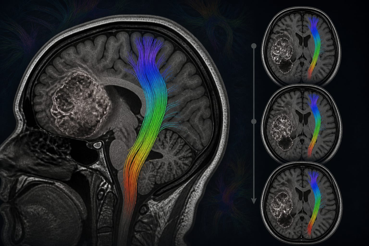
This retrospective, single-center study examined longitudinal changes in the contralesional corticospinal tract (CST) in 29 adults with frontal-lobe high-grade glioma, using DTI tractography at primary disease and first recurrence. The supplied article text describes the cohort, imaging, and tract-reconstruction methods but does not provide the study’s numerical results or direction of observed changes.
Background
Preserving motor pathways is an important consideration in glioma surgery, and preoperative DTI tractography is commonly used to delineate the CST in relation to a tumor. The contralesional CST is often used as a patient-specific comparator because it is distant from the lesion, an approach that assumes relative stability over time. Prior work cited in the article has reported diffusion abnormalities in contralateral normal-appearing white matter and serial white-matter alterations during high-grade glioma treatment. The study therefore assessed whether the contralesional CST changes between primary and recurrent disease, examining both tract properties and spatial distribution.
Data Highlights
Study feature
Details reported
Design and setting
Retrospective, single-center longitudinal study
Cohort
29 adults treated from 2013 through 2021
Eligibility
Frontal-lobe high-grade glioma, imaging-documented first recurrence, preoperative DTI at both assessments, and MRC strength of 5/5 at both assessments
Imaging interval to surgery
MRI obtained 1–7 days before surgery at each assessment
DTI acquisition
32 diffusion directions; b = 1000 s/mm²; 2-mm isotropic voxels
Tractography
Deterministic DTI-based reconstruction using an identical anatomically guided protocol at both assessments
Outcomes specified
Fractional anisotropy, tract length, volume, trajectory, and voxel-level distribution after spatial standardization
Key Findings
The study included 29 adults with frontal-lobe high-grade glioma and clinically imaging-documented first recurrence.
Participants were required to have MRC strength of 5/5 at both assessments, restricting the analysis to patients with preserved motor strength.
Preoperative MRI and DTI were obtained at both primary disease and recurrence, 1–7 days before surgery.
The contralesional CST was reconstructed at both assessments using deterministic DTI tractography and the same anatomically guided protocol.
The analysis was designed to assess both tract-level properties—fractional anisotropy, length, and volume—and spatial features, including trajectory and voxel-level distribution after spatial standardization.
The supplied article excerpt does not report the results of these comparisons; no direction or magnitude of longitudinal change can be stated from the provided material.
Clinical Implications
The study’s design directly examines the assumption that the contralesional CST remains stable between primary and recurrent high-grade glioma. However, the supplied material does not include outcome data or author conclusions, so it does not support a specific clinical recommendation about using the contralesional CST as a comparator.
Conclusion
This within-patient study was designed to characterize longitudinal contralesional CST changes using DTI tractography in high-grade glioma. The provided excerpt contains methods but not results, limiting conclusions about whether or how the tract changed.
by Bingzhen Li, Raimunde Liang, Ghaith Altawalbeh, Lisa Hoenikl, Eugen Ursu, Axel Schroeder, Haosu Zhang, Philipp Karschnia, Chiara Negwer, Bernhard Meyer, Arthur Wagner
Clinical trials continue to inform how physicians treat advanced prostate cancer. The American Society of Clinical Oncology (ASCO) develops guidelines to help oncologists evaluate new evidence and apply it to clinical practice
Antibody-drug conjugates (ADCs) have become an increasingly important part of treatment for several advanced cancers, pairing a tumor-targeting antibody with a cytotoxic payload.