Case Report: Carotid body paraganglioma in a 20-year-old woman: multimodal imaging, vessel-preserving surgical excision, and long-term clinical follow-up - Report - MDSpire
Advertisement
Case Report: Carotid body paraganglioma in a 20-year-old woman: multimodal imaging, vessel-preserving surgical excision, and long-term clinical follow-up
Clinical Report: A 20-Year-Old Female with Carotid Body Paraganglioma
Background
Carotid body paragangliomas are rare neuroendocrine tumors that typically present as painless neck masses. Their occurrence in young adults is uncommon.
Data Highlights
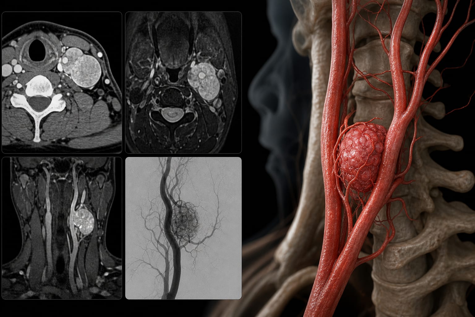
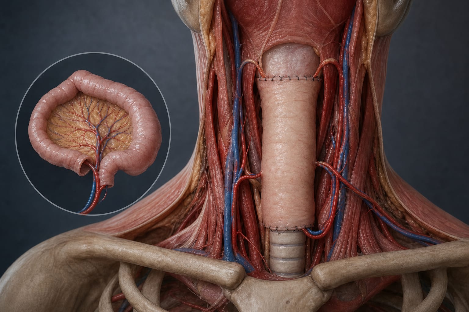
Multimodal imaging techniques including ultrasonography, CTA, MRI, and DSA were utilized for diagnosis. Surgical excision achieved grossly complete removal while preserving surrounding vascular structures.
Key Findings
The patient presented with a painless left lateral neck mass that had been enlarging over 5 years.
Imaging confirmed a hypervascular mass at the left carotid bifurcation.
Surgical excision was performed with preservation of the carotid arteries and cranial nerves.
Histopathology confirmed the diagnosis of carotid body paraganglioma.
Postoperative follow-up showed no recurrence-related symptoms or significant vascular abnormalities.
Clinical Implications
This case highlights the importance of multimodal imaging in the diagnosis and surgical planning of carotid body paragangliomas.
Conclusion
The case illustrates the management of carotid body paraganglioma in a young patient, emphasizing the role of comprehensive imaging and careful surgical technique.