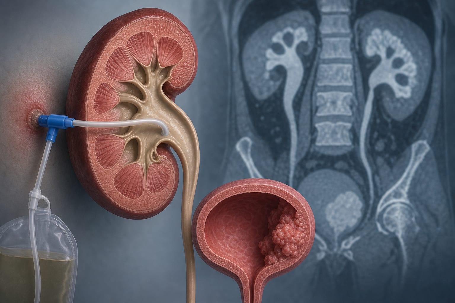
German S3 Guidelines on Diagnostic and Prognostic Assessment in Renal Cell Tumors
Overview
The German S3 Guidelines provide evidence-based recommendations for the diagnosis, staging, and prognostic evaluation of renal cell tumors, emphasizing advanced imaging techniques, biopsy strategies, and histopathological classification. These guidelines aim to optimize individualized treatment by integrating imaging, biopsy, and emerging molecular biomarkers.
Background
Renal cell tumors have seen evolving therapeutic options over the past two decades, including systemic therapies and surgical techniques. The increasing detection of small renal masses necessitates precise diagnostic and prognostic tools to guide treatment decisions. Imaging modalities such as CT and MRI, along with renal tumor biopsy and histopathological classification, play critical roles in diagnosis and staging. There is also a recognized need for molecular biomarkers to enhance diagnostic accuracy and prognostic stratification.
Data Highlights
Modality
Use
Accuracy/Performance
CT
Routine imaging for small RCC, staging including thorax
Staging accuracy 0.78-0.87; good for perirenal fat infiltration; limited for intrarenal infiltration
MRI
Preferred for caval thrombus, iodine allergy, brain metastases
Sensitivity 1.0 and specificity 0.83 for caval thrombus detection
Biopsy (Core)
Histopathological diagnosis, especially for uncertain lesions
Diagnostic yield and sensitivity ~99%; low morbidity; repeat biopsy recommended if negative but suspicion remains
Key Findings
High-resolution CT and MRI are essential for detecting and staging small renal cell carcinomas, with CT routinely used and MRI preferred for caval thrombus evaluation.
CT scans should include unenhanced, early arterial, and delayed phases with thin slices (2 mm) and 3D reconstructions for surgical planning.
Biopsy, particularly core biopsy, has a high diagnostic accuracy and is recommended for uncertain renal lesions, especially when active surveillance or ablation is considered.
Negative or indeterminate biopsy results require repeat biopsy or should be interpreted cautiously in the context of suspicious imaging findings.
Histopathological classification follows WHO and ISUP standards, with recognition of new RCC entities to improve diagnostic precision.
Emerging imaging techniques like diffusion and perfusion MRI show promise for grading but require further validation.
Clinical Implications
Clinicians should utilize high-resolution CT as the primary imaging modality for small renal tumors and reserve MRI for specific indications such as caval thrombus or contrast allergies. Core biopsy is a valuable tool to confirm diagnosis and guide management, particularly in ambiguous cases or when considering conservative approaches. Awareness of updated histopathological classifications supports accurate tumor typing and prognostication. Repeat biopsy should be considered when initial results are inconclusive but clinical suspicion remains high.
Conclusion
The German S3 Guidelines synthesize current evidence to recommend a multimodal diagnostic approach combining advanced imaging, biopsy, and histopathological classification to optimize the management of renal cell tumors. Continued research into molecular biomarkers and imaging techniques is needed to further individualize patient care.