Incisura groove depth is associated with reduced rotational fibular displacement after syndesmotic injury: cadaveric validation of an automated three-dimensional incisura morphometric pipeline - Report - MDSpire
Advertisement
Incisura groove depth is associated with reduced rotational fibular displacement after syndesmotic injury: cadaveric validation of an automated three-dimensional incisura morphometric pipeline
Clinical Report: Association of Incisura Groove Depth with Fibular Displacement
Overview
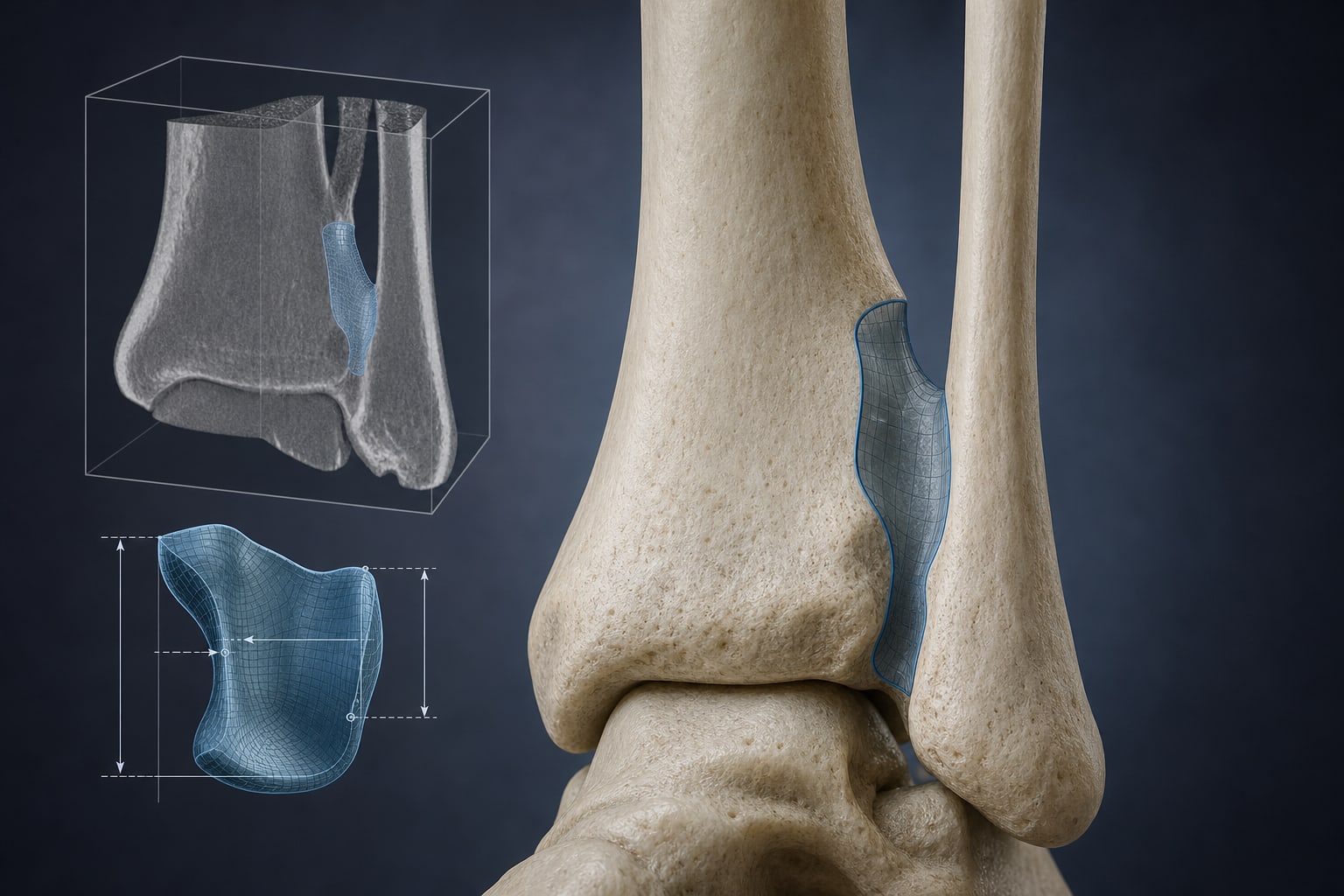
This study validates an automated 3D morphometric analysis of the tibial incisura, demonstrating that deeper incisura groove depths are associated with reduced fibular displacement following syndesmotic injury in a cadaveric model.
Background
The distal tibiofibular syndesmosis is crucial for ankle stability, and its disruption is common in athletic injuries. Accurate reduction of the fibula within the tibial incisura is essential for optimal recovery, yet malreduction remains a significant concern. Understanding the relationship between incisura morphology and fibular displacement is important.
Data Highlights
Parameter
Correlation Coefficient (r)
p-value
Mean groove depth
-0.48
0.004
Maximum groove depth
-0.46
0.006
Single-level incisura floor depth
-0.44
0.010
Total groove volume
-0.37
0.030
Key Findings
The automated toolbox demonstrated reproducibility across repeat CT acquisitions.
Bilateral reliability for morphometric outputs was good, with ICC values ≥ 0.75.
Four of six morphometric outputs were significantly negatively associated with fibular displacement magnitude.
The first principal component explained 85.4% of variance in the data.
Incisura morphology may influence surgical planning and injury prediction models.
Clinical Implications
The findings indicate that assessing incisura morphology could be relevant in the context of syndesmotic injuries.
Conclusion
The study provides a validated method for quantifying tibial incisura morphology, highlighting its potential role in understanding fibular displacement dynamics following syndesmotic injuries.