Development and validation of a predictive nomogram for leptomeningeal metastasis risk in NSCLC brain metastases: role of tumor location, driver mutations, and stereotactic radiosurgery - Report - MDSpire
Advertisement
Development and validation of a predictive nomogram for leptomeningeal metastasis risk in NSCLC brain metastases: role of tumor location, driver mutations, and stereotactic radiosurgery
Nomogram Predicting Leptomeningeal Metastasis Risk in NSCLC Brain Metastases
Overview
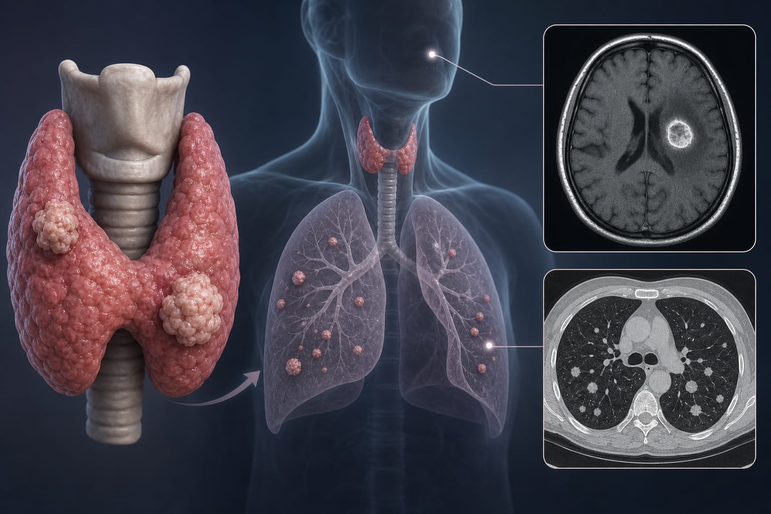
Leptomeningeal metastasis (LM) is a severe complication in NSCLC patients with brain metastases (BM), associated with poor prognosis. This study developed a nomogram incorporating tumor site, genetic mutations, and stereotactic radiosurgery to predict LM risk, aiding clinical decision-making.
Background
Leptomeningeal metastasis occurs in approximately 4–7% of NSCLC patients with CNS involvement and is linked to dismal survival outcomes. LM incidence is higher in patients harboring EGFR and ALK mutations, particularly prevalent in Asian populations. Metastatic spread to the leptomeninges may arise from multiple routes, including direct seeding from brain metastases, which are common precursors to LM. Surgical resection and radiotherapy have been implicated as potential risk factors for LM development.
Data Highlights
The retrospective cohort included 112 NSCLC patients with brain metastases: 56 who developed LM subsequently and 56 who did not develop LM until death. Patients with prior whole brain radiation therapy were excluded. Diagnosis of LM was confirmed by MRI and CSF cytology. Baseline clinical, radiological, and treatment characteristics at BM diagnosis were analyzed to identify predictors of LM development.
Key Findings
LM developed subsequent to BM in NSCLC patients, with 52.5–57.8% of LM cases having BM comorbidity.
EGFR and ALK mutations were associated with higher LM incidence (9.4% and 10.3%, respectively), more common in Asian populations.
Surgical resection of brain metastases increased risk of nodular-type LM, but pre-operative irradiation could mitigate this risk.
The nomogram developed integrates tumor site, genetic mutation status, and stereotactic radiosurgery to predict LM risk accurately.
Whole brain radiation therapy prior to LM diagnosis was excluded to avoid confounding effects.
Clinical Implications
The nomogram provides a practical tool for clinicians to stratify LM risk in NSCLC patients with brain metastases, facilitating personalized treatment planning. Awareness of genetic mutation status and prior treatments such as stereotactic radiosurgery can guide surveillance intensity and therapeutic decisions. Pre-operative irradiation may reduce LM risk in patients undergoing surgical resection of brain metastases.
Conclusion
This study highlights key predictors of leptomeningeal metastasis in NSCLC patients with brain metastases and presents a validated nomogram to aid clinical risk assessment. Incorporating tumor characteristics and treatment factors can optimize management strategies to potentially improve patient outcomes.
References
Zhen et al. 2020 -- Association of Brain Metastases with Leptomeningeal Metastasis in NSCLC
Yin et al. 2019 -- Brain Metastases as a Risk Factor for Leptomeningeal Metastasis
Guangdong Sanjiu Brain Hospital Ethics Committee 2021 -- Retrospective Study on LM Prediction