Forecasting Lesion Recovery in Acute Cerebral Ischemia Using ADC Thresholds
Overview
This study evaluated the predictive value of voxel-based apparent diffusion coefficient (ADC) thresholds from diffusion-weighted MRI (DWI) for lesion reversal in acute ischemic stroke patients undergoing endovascular therapy (EVT). Findings indicate that a single ADC threshold cannot reliably distinguish salvageable from unsalvageable tissue, and a lower ADC boundary exists below which lesion reversal is unlikely.
Background
Accurate assessment of ischemic lesion extent is critical in acute stroke management to guide reperfusion therapies such as EVT. Diffusion-weighted imaging (DWI) is sensitive to early ischemic changes, but DWI hyperintensities do not always represent irreversible infarction, as lesion reversal can occur with reperfusion. The apparent diffusion coefficient (ADC) is used to differentiate infarct core from penumbra, but physiological and technical variability complicates establishing universal ADC thresholds. Understanding ADC thresholds is essential for accurate stroke imaging interpretation and clinical decision-making.
Data Highlights
Parameter
Value/Description
ADC threshold commonly cited
620 × 10⁻⁶ mm²/s
ADC voxel exclusion criteria
Above 760 × 10⁻⁶ mm²/s or below 200 × 10⁻⁶ mm²/s
Inter-rater Dice coefficient for segmentation
0.852 (95% CI: 0.834–0.880)
Time window for recanalization
Within 2 hours after MRI
Pre-EVT DWI lesion volume inclusion
Greater than 1 mL
Key Findings
DWI lesion reversal (DWI-R) occurs predominantly in patients with rapid and successful recanalization by EVT.
A single voxel-based ADC threshold is insufficient to reliably distinguish salvageable from unsalvageable ischemic tissue.
Physiological variability in ADC values across brain regions and technical factors limit the universality of ADC thresholds.
There exists a lower ADC boundary below which lesion reversal is negligible, indicating irreversible infarction.
Segmentation reliability was high with a Dice coefficient of 0.852, supporting robustness of lesion delineation.
Strict inclusion criteria (e.g., recanalization within 2 hours, lesion volume >1 mL) were applied to minimize confounding factors.
Clinical Implications
Clinicians should exercise caution when using fixed ADC thresholds to define infarct core, as over-reliance may exclude patients with potentially salvageable tissue from reperfusion therapies. Incorporating knowledge of ADC variability and considering time to recanalization can improve interpretation of DWI and ADC maps. This nuanced approach supports better patient selection for EVT and may optimize functional outcomes.
Conclusion
The study underscores the limitations of a single ADC threshold to predict lesion recovery in acute ischemic stroke. A tailored interpretation of ADC values, accounting for physiological and technical factors, is essential to guide reperfusion treatment decisions effectively.
References
Olsen et al. 2024 -- Forecasting Lesion Recovery in Acute Cerebral Ischemia Using ADC Thresholds
by Thor Håkon Skattør, Atle Bjørnerud, Terje Nome, Kine Mari Bakke, Brian Anthony Enriquez, Ingrid Digernes, Cecilie Mørck Offersen, Mona Kristiansen Beyer, Geir Ringstad, Anne Hege Aamodt
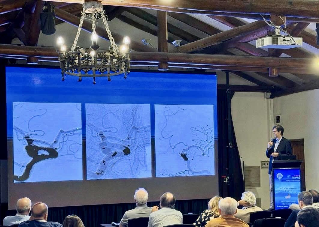
Guilherme Dabus, M.D., co-director of interventional neuroradiology at Baptist Health Miami Neuroscience Institute, served as a guest professor and invited speaker at the GSANIT (Grupo Sudamericano de Neurorradiología Intervencionista y Terapeutica) in Santa Cruz, Chile,