STIR or T2-Dixon? A false dilemma in musculoskeletal MRI - Report - MDSpire

STIR or T2-Dixon? A false dilemma in musculoskeletal MRI

  • By

  • Teodoro Martín-Noguerol

  • Oscar L. Casado-Verdugo

  • Antonio Luna

  • July 1, 2026

  • 0 min

Share

Clinical Report: STIR versus T2-Dixon in Musculoskeletal MRI Techniques

Background

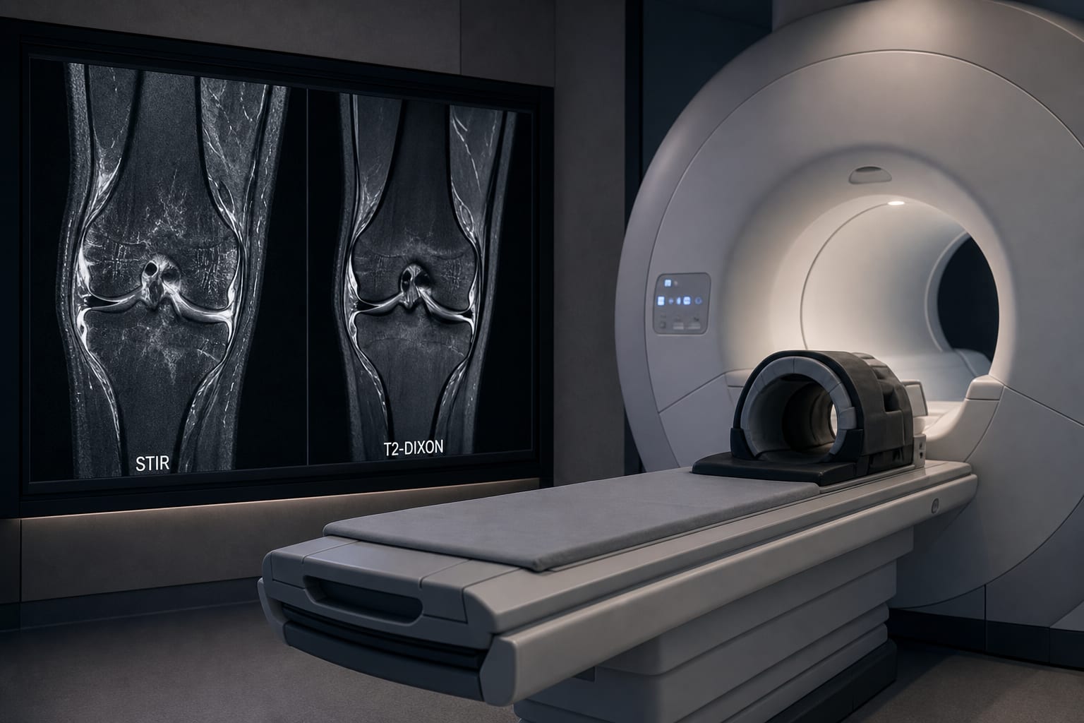
The choice between STIR and T2-Dixon sequences is critical in musculoskeletal MRI, particularly for bone marrow assessment. STIR has been a trusted technique since the 1980s, known for its excellent fat suppression and sensitivity to edema. In contrast, T2-Dixon, which emerged later, offers unique advantages but has faced hurdles in consistent clinical implementation due to technological and workflow challenges.

Data Highlights

No numerical data available in the article.

Key Findings

  • STIR provides excellent fat suppression and is less vulnerable to magnetic field inhomogeneities.
  • T2-Dixon sequences were introduced later due to technological limitations and inconsistent implementation across different MRI systems.
  • Radiologists have raised concerns about the quality and reliability of T2-Dixon images compared to STIR.
  • Despite initial challenges, T2-Dixon has shown potential advantages in generating multiple image sets from a single acquisition.
  • Recent studies indicate that T2-Dixon may offer benefits in specific clinical scenarios, such as post-contrast imaging.

Clinical Implications

Radiologists should consider the strengths and limitations of both STIR and T2-Dixon sequences when selecting imaging protocols for musculoskeletal evaluations. Understanding the historical context and technological advancements can aid in optimizing MRI practices.

Conclusion

The ongoing evolution of MRI techniques necessitates a careful assessment of STIR and T2-Dixon sequences to enhance diagnostic accuracy in musculoskeletal imaging.

Related Resources & Content

  1. European Radiology, 2023 -- Comparative Analysis of STIR and T2 Dixon Sequences in Pediatric Whole-Body MRI for Identifying and Assessing High Signal Bone Marrow Alterations
  2. European Radiology, 2024 -- Creating High-Resolution Spinal STIR and T1-weighted Images from Low-Resolution Axial Dixon and T2-weighted FSE Sequences
  3. European Radiology, 2025 -- Impact of Imaging Techniques on Fat Fraction Measurement for Evaluating Bone Marrow in Metastatic Prostate Cancer
  4. La radiologia medica, 2026 -- Whole-body magnetic resonance imaging in rheumatology: advancements, key applications and future perspectives
  5. Radiology, 2026 -- How I Do It: Using the Dixon Method and Fat-Water Imaging in Musculoskeletal MRI
  6. European Radiology Experimental, 2025 -- Quantitative MRI Dixon signal drop and fat fraction for differentiating bone marrow lesions: a two-center prospective analysis
  7. European Radiology — Evaluating the Diagnostic Efficacy of Dual-Energy CT Virtual Monochromatic Imaging for Supraspinatus Tendon Injuries Compared to Conventional CT and MRI Techniques
  8. Whole-body magnetic resonance imaging in rheumatology: advancements, key applications and future perspectives | La radiologia medica | Springer Nature Link
  9. How I Do It: Using the Dixon Method and Fat-Water Imaging in Musculoskeletal MRI
  10. Quantitative MRI Dixon signal drop and fat fraction for differentiating bone marrow lesions: a two-center prospective analysis | European Radiology Experimental | Springer Nature Link

Original Source(s)

Related Content