Subtype-specific sonographic signatures and clinicopathological features of metaplastic breast cancer: a 50-case cohort study
By
Boxiong Wei
Zijing Fu
Yuhong Shao
Xiuming Sun
Luzeng Chen
July 2, 2026
Clinical Report: Sonographic Characteristics and Clinical-Pathological Aspects of Metaplastic Breast Cancer
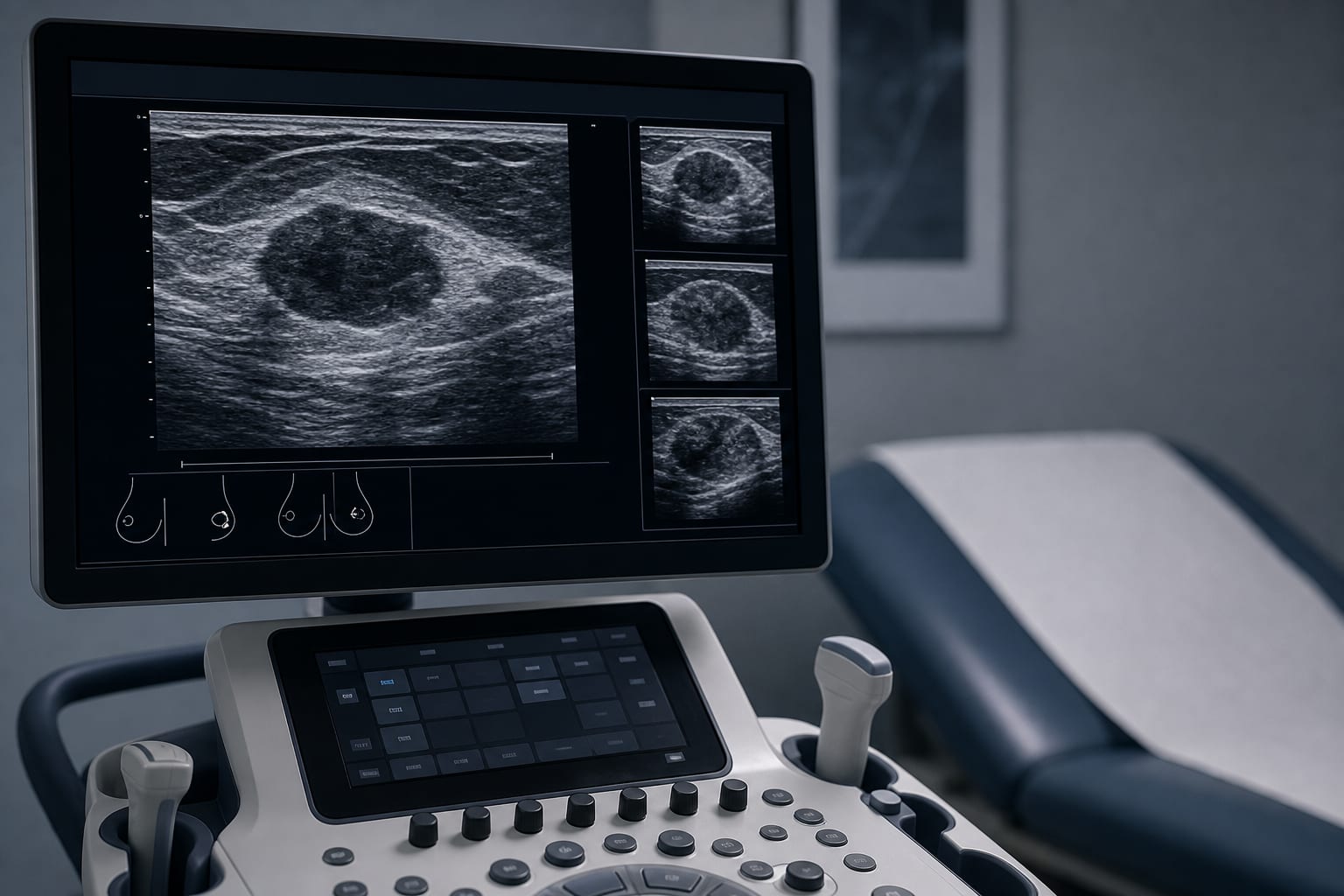
Overview This study investigates the sonographic features and clinicopathological characteristics of metaplastic breast cancer (MBC) across different subtypes.
Background Metaplastic breast cancer (MBC) is a rare and aggressive form of breast cancer, accounting for less than 1% of all breast carcinomas. It is often associated with poor prognosis and low response rates to conventional chemotherapy. Accurate diagnosis is important, as MBC can exhibit atypical imaging features that may lead to misinterpretation.
Data Highlights Subtype Sonographic Features Median Tumor Size (cm) Axillary Lymph Node Metastasis Rate (%) Spindle Cell Pseudo-benign appearance, circumscribed margins (60.0%), oval/round shape (46.7%) 3.1 22.0 Squamous Cell Heterogeneous echogenicity (83.3%), complex cystic-solid components (75.0%) N/A N/A Mixed Type Heterogeneous echogenicity (73.9%), complex cystic-solid components (56.5%) N/A N/A
Key Findings The spindle cell subtype of MBC often presents with a pseudo-benign sonographic phenotype. Spindle cell tumors have significantly smaller median dimensions compared to squamous and mixed types. Squamous cell and mixed-type tumors frequently exhibit heterogeneous echogenicity and complex cystic-solid components. The overall axillary lymph node metastasis rate is 22%. There is a notable size-node dissociation pattern in MBC. Clinical Implications Recognizing the distinct sonographic features of MBC subtypes is essential for accurate diagnosis.
Conclusion The study highlights the significant heterogeneity of MBC.
Related Resources & Content
Frontiers in Oncology, 2026 -- Navigating the Diagnostic Pitfalls of Cystic and Solid Breast Masses: A Comparative Case Report of Metaplastic Carcinoma and Benign Lesions Frontiers in Oncology, 2026 -- Metaplastic breast carcinoma with osteosarcomatous differentiation: a case report Frontiers in Oncology, 2026 -- Multimodal imaging features of primary breast epithelial–myoepithelial carcinoma: a case report and literature review Cytologic analysis of metaplastic breast carcinoma: Review of 66 cases diagnosed at the Institut Curie | American Journal of Clinical Pathology, 2023 Multimodal Imaging Features and Prognosis of Metaplastic Breast Carcinoma - PubMed, 2024 Frontiers in Oncology — Case Report: Granular cell tumor of the axillary tail mimicking node-positive breast carcinoma — two cases with imaging–pathology correlation Cytologic analysis of metaplastic breast carcinoma: Review of 66 cases diagnosed at the Institut Curie | American Journal of Clinical Pathology | Oxford Academic Multimodal Imaging Features and Prognosis of Metaplastic Breast Carcinoma - PubMed A Systematic Review With Individual Patient Data Meta-analysis on Characteristics and Outcomes of Patients With Metaplastic Breast Carcinoma - ScienceDirect