MRI Assessment of Liver and Abdominal Fat Volumes Before and After Bariatric Surgery
Overview
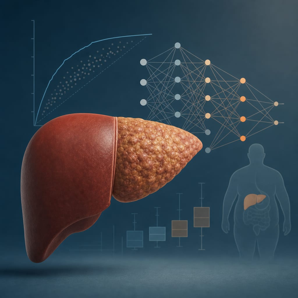
This study evaluated changes in liver, visceral, and subcutaneous fat volumes in morbidly obese patients undergoing bariatric surgery using advanced MRI techniques. Significant reductions in fat volumes were observed postoperatively, alongside changes in liver biomarkers, highlighting MRI as a valuable non-invasive tool for monitoring fat distribution and liver health.
Background
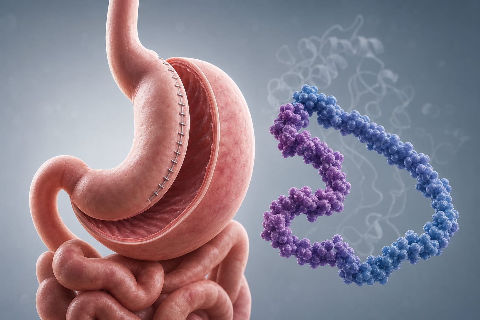
Obesity prevalence is rising globally, with fatty liver disease closely linked to increased BMI and risk of progression to severe liver conditions such as NASH and cirrhosis. Bariatric surgery is the most effective treatment for morbid obesity, leading to weight loss and reduction in intrahepatic and abdominal fat, which improves metabolic and cardiovascular outcomes. Liver biopsy remains the gold standard for assessing liver fat but is invasive and less feasible in morbidly obese patients, prompting the need for non-invasive imaging methods like MRI. Fat–water MRI techniques enable accurate quantification of fat content in liver and adipose tissues, facilitating safe monitoring before and after bariatric surgery.
Data Highlights
Parameter
Preoperative Mean (±SD)
Postoperative Timepoints
BMI
40.6 ± 1.0 kg/m2
Measured at 3, 6, 12, and 24 months post-surgery
Subjects
16 morbidly obese patients (2 male, 14 female)
12 healthy lean controls (12 male)
Age
34.4 ± 2.9 years (range 19–54)
24.8 years mean in controls
Imaging
Siemens 3T MAGNETOM Prisma MRI
Two-point Dixon and T2*-IDEAL sequences for fat quantification
Key Findings
MRI enabled precise quantification of liver fat percentage, total liver volume, visceral adipose tissue (VAT), and subcutaneous adipose tissue (SAT) in morbidly obese patients.
Significant reductions in liver fat and abdominal fat volumes were observed at multiple postoperative timepoints (3, 6, 12, and 24 months) following bariatric surgery.
Liver biomarkers including liver fatty acid-binding proteins (L-FABP), Fetuin A, and M30 were measured and showed dynamic changes post-surgery, indicating metabolic improvements.
The MRI sequences used allowed rapid acquisition during single breath holds, minimizing motion artifacts and improving data quality.
Non-invasive MRI fat quantification provides a safe alternative to liver biopsy for monitoring fatty liver disease progression and response to bariatric surgery in morbidly obese patients.
Clinical Implications
MRI-based fat quantification offers a reliable, non-invasive method to monitor liver and abdominal fat changes in morbidly obese patients undergoing bariatric surgery, circumventing the risks and limitations of liver biopsy. Regular MRI assessments can guide clinical management by tracking fat reduction and liver health improvements, potentially aiding in early detection of fatty liver disease progression or resolution. Incorporation of liver biomarkers alongside imaging may enhance understanding of metabolic changes post-surgery.
Conclusion
Advanced MRI techniques effectively quantify liver and abdominal fat volumes, demonstrating significant fat reduction after bariatric surgery in morbidly obese patients. This non-invasive approach holds promise for routine monitoring of fatty liver disease and metabolic improvements in this population.
References
Obesity and Fatty Liver Disease Epidemiology and Pathophysiology
Bariatric Surgery and Metabolic Outcomes in Morbid Obesity
MRI Techniques for Fat Quantification in Liver and Adipose Tissue
by Anne Christin Meyer-Gerspach, Ralph Peterli, Michael Moor, Philipp Madörin, Andreas Schötzau, Diana Nabers, Stefan Borgwardt, Christoph Beglinger, Oliver Bieri, Bettina K. Wölnerhanssen