Shallow and bullous retinal pigment epithelium elevations in age-related macular degeneration - Report - MDSpire
Coming Soon: Introducing MDSpire News. Learn more
Conexiant’s news site is now MDSpire News. Learn more

Elevations of the Retinal Pigment Epithelium in Age-Related Macular Degeneration: Shallow and Bullous Forms

  • By

  • Jost B. Jonas

  • Songhomitra Panda-Jonas

  • Jie Xue

  • Rahul A. Jonas

  • Ya Xing Wang

  • September 25, 2026

Share

Elevations of the Retinal Pigment Epithelium in Age-Related Macular Degeneration

Overview

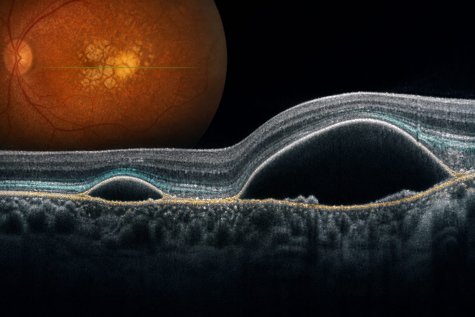
This study investigates the prevalence and associations of retinal pigment epithelium (RPE) elevations and detachments in a population-based cohort. It classifies these elevations into shallow and bullous forms.

Background

RPE elevations and detachments are significant features in the morphologic spectrum of AMD, which can lead to visual impairment. This study aims to address limitations in previous research by including a larger, population-based cohort.

Data Highlights

No numerical data or trial data provided in the source material.

Key Findings

  • RPE elevations can be classified as flat RPE elevations (RPEEs) or bullous RPE detachments (RPEDs).
  • Shallow and bullous RPE elevations are associated with a higher risk of AMD progression.
  • The study cohort included participants without macular disorders.
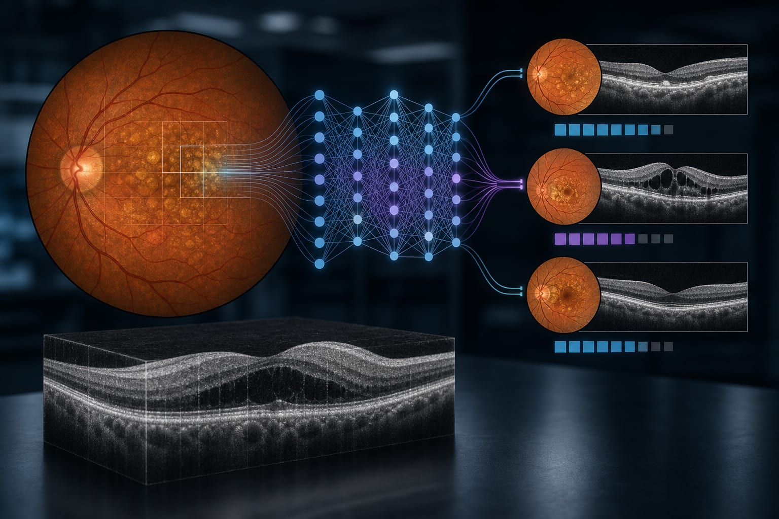
  • OCT imaging was utilized to define and classify RPE elevations and detachments.
  • Assessment included additional macular findings such as intraretinal hyperreflective foci and abnormalities of the outer nuclear layer.

Clinical Implications

The classification of RPE elevations is essential for understanding the risk of AMD progression. Clinicians should consider these findings in the context of patient assessments to better manage AMD.

Conclusion

This study provides insights into the prevalence and classification of RPE elevations in AMD.

Related Resources & Content

  1. Retinal Physician, 2018 -- Treatment of Pigment Epithelial Detachments in Age-Related Macular Degeneration
  2. Retinal Physician, 2012 -- Treatment of Pigment Epithelial Detachments in Age-related Macular Degeneration
  3. Retinal Physician, 2017 -- Dry AMD Masquerade Syndromes
  4. Ophthalmology Management, 2017 -- 'Real world' OCT: Subtle findings, critical implications
  5. Graefe's Archive for Clinical and Experimental Ophthalmology -- OCTA in age-related macular degeneration: consensus on practical guidelines for optimal imaging strategies
  6. Ophthalmology, 2025 -- Age-Related Macular Degeneration Preferred Practice Pattern®
  7. PubMed -- Intravitreal Anti-Vascular Endothelial Growth Factor Agents in Patients with Neovascular Age-Related Macular Degeneration and Retinal Pigment Epithelial Tear: A Systematic Review and Meta-Analysis
  8. OCTA in age-related macular degeneration: consensus on practical guidelines for optimal imaging strategies across different clinical scenarios | Graefe's Archive for Clinical and Experimental Ophthalmology | Springer Nature Link
  9. Age-Related Macular Degeneration Preferred Practice Pattern® - Ophthalmology
  10. Intravitreal Anti-Vascular Endothelial Growth Factor Agents in Patients with Neovascular Age-Related Macular Degeneration and Retinal Pigment Epithelial Tear: A Systematic Review and Meta-Analysis - PubMed

Original Source(s)

Related Content