Removal of Implantable Collamer Lenses Due to Elevated Intraocular Pressure in a Patient with Severe Hyperopia: A Case Study
-
By
-
Liuqing Cui
-
Karl Mercieca
-
Leonie Bourauel
-
March 24, 2026
Removal of Implantable Collamer Lenses for Elevated IOP in Severe Hyperopia
Overview
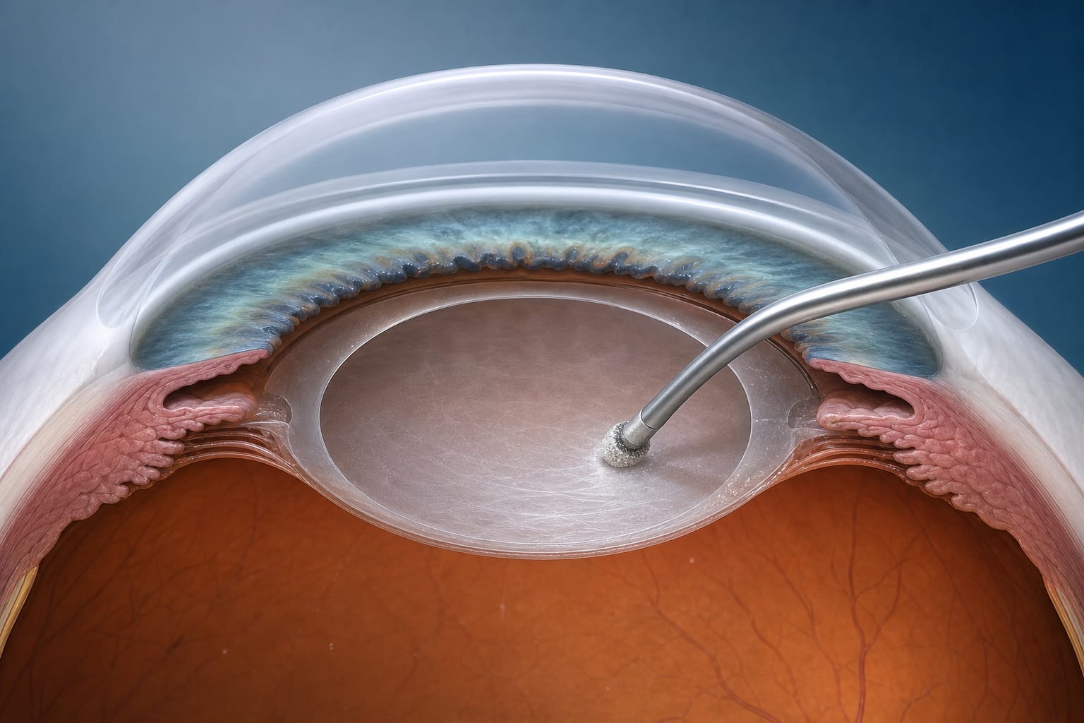
A 38-year-old woman with severe hyperopia developed elevated intraocular pressure (IOP) 15 years after implantable collamer lens (ICL) implantation. Despite medical therapy, IOP control was inadequate, leading to bilateral ICL explantation combined with cataract surgery, resulting in improved IOP and visual acuity.
Background
Posterior chamber phakic intraocular lens (PC pIOL) implantation is a recognized surgical option for correcting high hyperopia, preserving corneal anatomy and providing stable refractive outcomes. However, long-term complications such as elevated intraocular pressure, secondary glaucoma, and cataract formation can occur, especially with ICLs lacking a central aperture. This case study highlights the management challenges and surgical outcomes in a patient with bilateral ICLs who developed IOP decompensation 15 years post-implantation.
Data Highlights
| Parameter | Right Eye (OD) | Left Eye (OS) |
|---|---|---|
| Initial IOP (mmHg) | 22 | 22 |
| IOP spikes without medication (since 2021) | Up to 30 | Up to 30 |
| Post-explantation IOP at 1 month (mmHg) | 17 (with dorzolamide) | 15 (without medication) |
| Post-explantation IOP at 6 months (mmHg) | 13 (with dorzolamide) | 14 (with dorzolamide) |
| Best-corrected visual acuity (BCVA) pre-surgery | 20/20 | 20/20 |
| BCVA 1 day post-surgery | 20/40 | 20/63 |
| BCVA 1 week post-surgery | 20/16 | 20/80 |
| BCVA 1 month post-surgery | 20/16 | 20/20 |
Key Findings
- Patient developed bilateral elevated IOP up to 30 mmHg 15 years after ICL implantation for high hyperopia.
- Medical therapy with bimatoprost, dorzolamide, and clonidine was insufficient to control IOP spikes.
- Slit-lamp and gonioscopy revealed well-positioned ICLs without central port, partial angle closure, peripheral anterior synechiae, and iris adhesions.
- Combined ICL explantation and phacoemulsification with posterior chamber IOL implantation were performed in both eyes.
- Postoperative course showed improved IOP control (14–17 mmHg) with reduced medication and restoration of visual acuity to 20/20 in both eyes.
- Post-surgical inflammation was managed successfully with topical and subconjunctival steroids.
Clinical Implications
Long-term monitoring of patients with ICLs, especially those without a central port, is essential due to the risk of late-onset elevated IOP and angle closure. In cases of inadequate medical IOP control and progressive angle compromise, combined explantation of the ICL and cataract surgery can effectively restore IOP control and visual function. Careful surgical planning and postoperative anti-inflammatory management are critical to optimize outcomes.
Conclusion
This case underscores the potential for late IOP elevation after ICL implantation in severe hyperopia and demonstrates that combined ICL removal with cataract surgery can successfully manage elevated IOP and preserve vision. Ongoing surveillance and individualized treatment strategies remain paramount in these patients.
Related Resources & Content
- 1 -- Safety and efficacy of PC pIOL implantation in high hyperopia
- 2 -- Complications associated with ICLs without central aperture
Based on findings from:
Removal of Implantable Collamer Lenses Due to Elevated Intraocular Pressure in a Patient with Severe Hyperopia: A Case Study
Liuqing Cui, Karl Mercieca, Leonie Bourauel. Bmc Ophthalmology, 2026.
https://link.springer.com/article/10.1186/s12886-026-04757-1
This content is an AI-generated, fully rewritten summary based on a published scholarly article. It does not reproduce the original text and is not a substitute for the original publication. Readers are encouraged to consult the source for full context, data, and methodology.