CT-based habitat imaging integrated with radiomics and clinicopathology for noninvasive prediction of microvascular invasion in hepatocellular carcinoma - Report - MDSpire

CT-based habitat imaging integrated with radiomics and clinicopathology for noninvasive prediction of microvascular invasion in hepatocellular carcinoma

  • By

  • Shuangxi Chen

  • Xushuang Qin

  • Shanni Dong

  • Xiaoshu Zhu

  • Yang Liu

  • Jun Chen

  • Ruizhong Ye

  • Li Zhu

  • May 22, 2026

  • 0 min

Share

Integration of CT Habitat Imaging with Radiomics for MVI Prediction in HCC

Overview

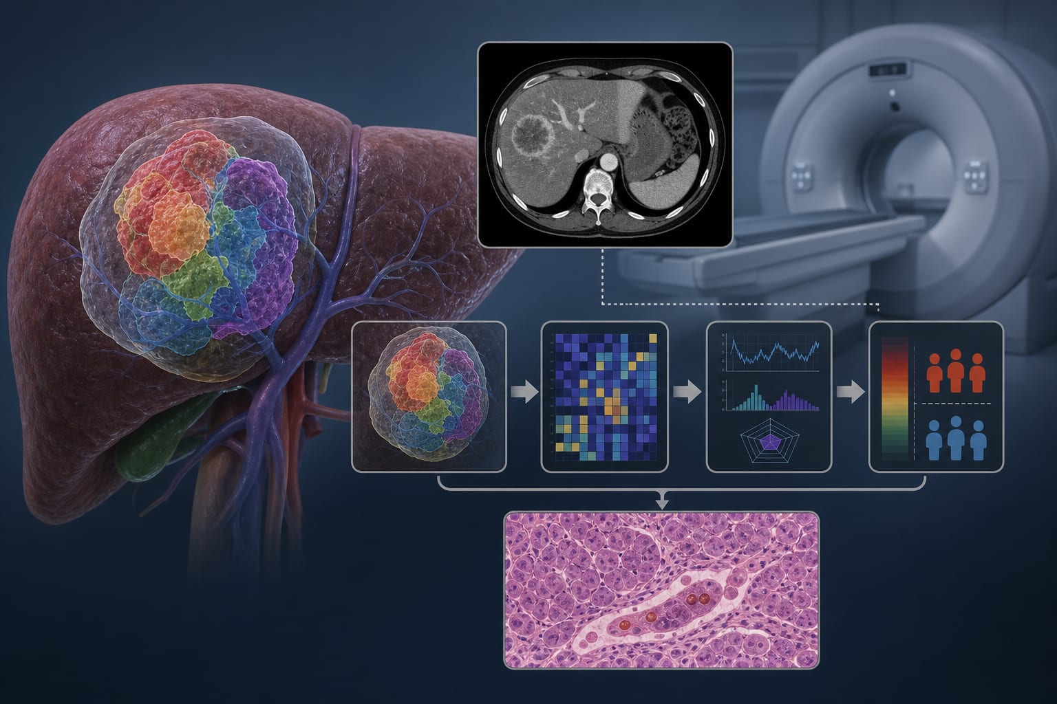
This study developed a CT-based habitat imaging model that integrates intratumoral microenvironment analysis with radiomics and clinicopathological data to predict microvascular invasion (MVI) in hepatocellular carcinoma (HCC). The model demonstrated superior predictive performance compared to conventional methods, highlighting its potential for noninvasive preoperative risk stratification.

Background

Hepatocellular carcinoma (HCC) is a leading cause of cancer-related mortality, with microvascular invasion (MVI) being a critical factor influencing prognosis and treatment decisions. Current methods for determining MVI are invasive and limited in capturing tumor heterogeneity. This study addresses the need for a reliable, noninvasive predictive tool that can enhance clinical decision-making in HCC management.

Data Highlights

ModelAUC (Training Set)AUC (Validation Set)
Combined Model0.8620.814
Clinical ModelNot reportedNot reported
Radiomics-Only ModelNot reportedNot reported

Key Findings

  • The combined model achieved an AUC of 0.862 in the training set and 0.814 in the validation set.
  • Calibration curves indicated good agreement with a Hosmer–Lemeshow P value of 0.60.
  • Decision curve analysis showed net benefit at thresholds of 15%–65%.
  • The combined model significantly outperformed both the clinical model and the radiomics-only model (P < 0.05).
  • Habitat analysis effectively quantified intratumoral heterogeneity, enhancing predictive accuracy for MVI.

Clinical Implications

The CT-based habitat imaging model provides a noninvasive method for preoperative MVI risk stratification in HCC patients, potentially guiding surgical planning and postoperative management. Its integration of radiomics and clinical data may improve individualized treatment approaches and patient outcomes.

Conclusion

The study presents a promising noninvasive tool for predicting microvascular invasion in HCC, which could significantly impact clinical practice by refining preoperative risk assessment and treatment strategies.

Related Resources & Content

  1. Frontiers in Oncology, 2026 -- Habitat and peritumoral CT radiomics accurately predict early treatment response to hepatic arterial infusion chemotherapy combined with tyrosine kinase inhibitors and programmed death‑1 inhibitors in unresectable hepatocellular carcinoma
  2. Frontiers in Medicine, 2026 -- Prediction of the efficacy after the first transarterial chemoembolization in hepatocellular carcinoma using CT radiomics combined with inflammatory composite indicators
  3. European Radiology, 2025 -- Analysis of 3D Fractal Dimensions in CT Imaging for Predicting Microvascular Invasion in Hepatocellular Carcinoma
  4. EASL Clinical Practice Guidelines on the management of hepatocellular carcinoma - PubMed
  5. European Radiology — Creation and assessment of a radiopathomics model for forecasting liver metastases in colorectal cancer patients
  6. Why MVI prediction matters now
  7. MRI-based Intra- and Peritumoral Heterogeneity in Hepatocellular Carcinoma for Microvascular Invasion Prediction and Prognostic Risk Stratification | Radiology: Imaging Cancer

Original Source(s)

Related Content