Effect of RAS inhibitors on retinal microvasculature in DKD using AI-based ultra-wide-field images - Report - MDSpire

Effect of RAS inhibitors on retinal microvasculature in DKD using AI-based ultra-wide-field images

  • By

  • Yan Liu

  • Xinyu Zhao

  • Haiyun Wang

  • Jie Ma

  • Peng Xia

  • Xingwang Gu

  • Youxin Chen

  • Limeng Chen

  • June 26, 2026

  • 0 min

Share

Clinical Report: Impact of RAS Inhibitors on Retinal Microvascular Health

Overview

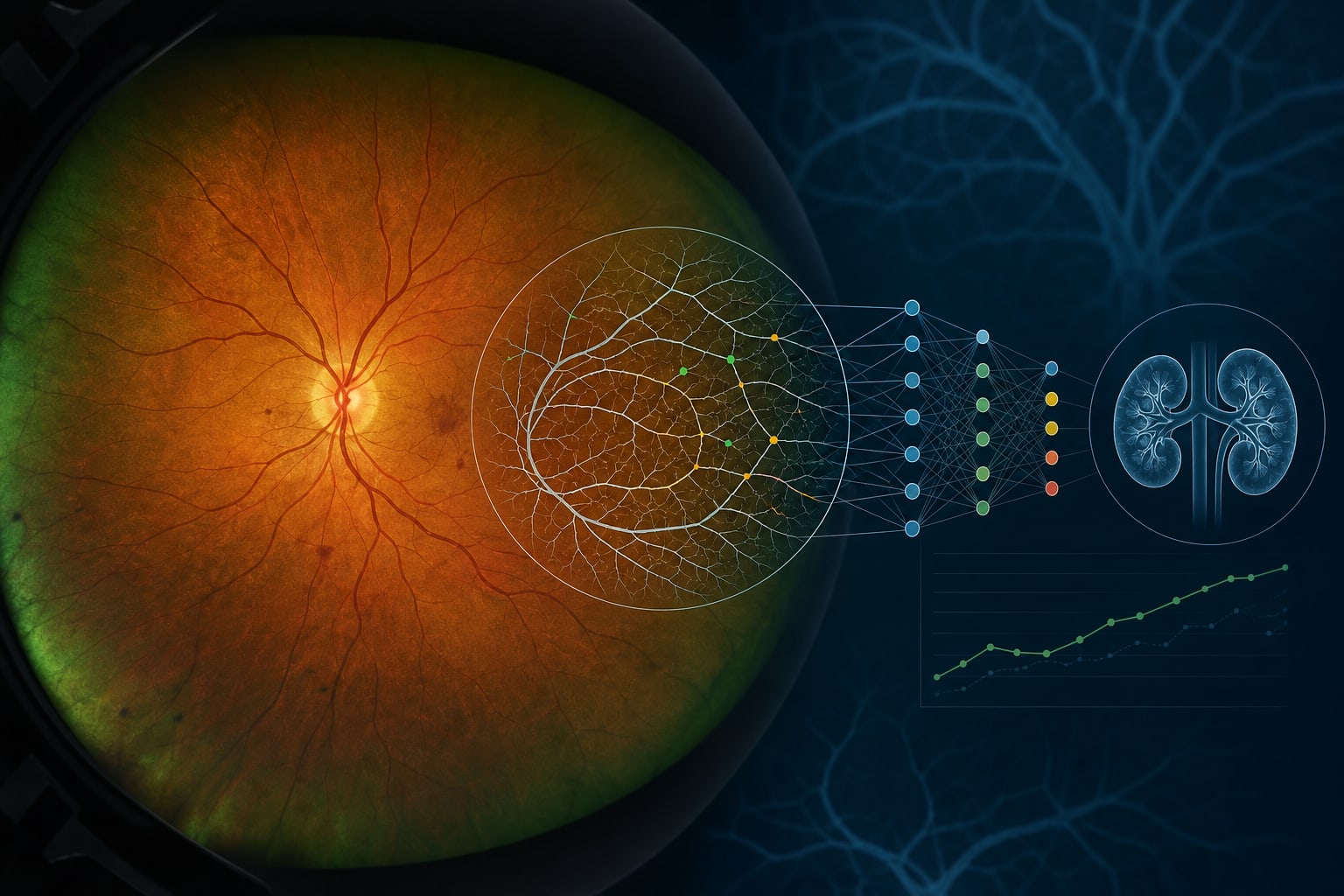
This pilot study evaluates the short-term effects of renin-angiotensin system inhibitors (RASi) on retinal microvascular parameters in patients with diabetic kidney disease (DKD). Significant decreases in venous fractal dimension and tortuosity were observed after 12 weeks of RASi therapy.

Background

Diabetic kidney disease (DKD) and diabetic retinopathy (DR) are significant microvascular complications of diabetes mellitus, often occurring together. RAS inhibitors have been shown to provide protective effects on both DKD and DR, yet their specific impact on retinal microvascular parameters remains unclear.

Data Highlights

ParameterBaselinePost-Treatmentp-value
Venous DfNot specifiedDecreased0.0389
Venous TORTNot specifiedDecreased0.0496

Key Findings

  • 21 out of 27 patients (77.8%) were male, with a mean age of 55.7 years.
  • 15 patients (55.6%) had diabetic retinopathy (DR).
  • Significant decreases in venous fractal dimension (Df) and tortuosity (TORT) were observed after 12 weeks of RASi treatment.
  • No significant changes were noted in parameters derived from the central retinal region.
  • Changes in retinal microvascular parameters correlated with alterations in clinical renal indicators.

Clinical Implications

The findings indicate specific alterations in peripheral retinal venous parameters after RASi therapy.

Conclusion

This study highlights the impact of RASi on retinal microvascular health in DKD patients.

Related Resources & Content

  1. Retinal Physician, Ultrawidefield Imaging for Diabetic Retinopathy, 2019 -- Extending the edge of discovery.
  2. Frontiers in Medicine, Artificial intelligence-based quantification of retinal microvascular biomarkers from fundus photography of chronic kidney disease: a case-control study.
  3. npj Digital Medicine, Unsupervised 3D Mapping and Analysis of Retinal Microvasculature Using RADAR.
  4. American Diabetes Association, Standards of Care in Diabetes—2026 -- Chronic Kidney Disease and Risk Management.
  5. KDIGO, 2026 Clinical Practice Guideline for Diabetes and Chronic Kidney Disease (CKD).
  6. Retinal Physician — Clinical Trials Provide Rationale for Use of Anti-VEGF Treatment Earlier in Diabetic Retinopathy
  7. Diabetic Retinopathy Preferred Practice Pattern® - Ophthalmology.
  8. 11. Chronic Kidney Disease and Risk Management: Standards of Care in Diabetes—2026 | Diabetes Care | American Diabetes Association
  9. KDIGO 2026 Clinical Practice Guideline for Diabetes and Chronic Kidney Disease (CKD)
  10. Diabetic Retinopathy - Endotext - NCBI Bookshelf
  11. Renal and Retinal Effects of Enalapril and Losartan in Type 1 Diabetes | New England Journal of Medicine
  12. Effect of candesartan on progression and regression of retinopathy in type 2 diabetes (DIRECT-Protect 2): a randomised placebo-controlled trial - ScienceDirect
  13. (PDF) Effect of candesartan on prevention (DIRECT-Prevent 1) and progression (DIRECT-Protect 1) of retinopathy in type 1 diabetes: randomised, placebo-controlled trials
  14. The Randomized Olmesartan and Diabetes Microalbuminuria Prevention (ROADMAP) observational follow-up study: benefits of RAS blockade with olmesartan treatment are sustained after study discontinuation - PubMed
  15. Association of Diabetic Lesions and Retinal Nonperfusion Using Widefield Multimodal Imaging - ScienceDirect
  16. Impact of Anatomically Defined Retinal Zones on Ultrawide-Field Fluorescein Angiography Nonperfusion Measurements in Diabetic Retinopathy - PubMed
  17. Automated Machine Learning for Predicting DR Progression From Ultra-Widefield Retinal Images
  18. Screening chronic kidney disease through deep learning utilizing ultra-wide-field fundus images
  19. Macular Perfusion Deficits on OCTA Correlate with Non-perfusion on Ultrawide-field FA in Diabetic Retinopathy - PMC

Original Source(s)

Related Content