Impact of Dural Attachment on Residual Tumor Progression in WHO Grade 1 Meningiomas
Overview
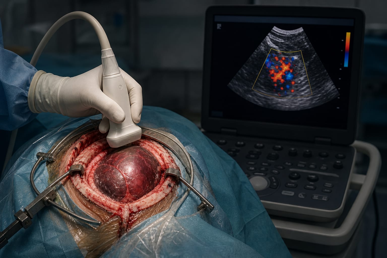
This study evaluates how dural attachment and MRI signal intensity influence the progression of residual WHO grade 1 meningioma tumors following subtotal resection. Findings suggest that the extent of dural contact and tumor signal characteristics are important predictors of remnant growth.
Background
Meningiomas are the most common intracranial tumors, often treated surgically. Complete resection is not always feasible, resulting in residual tumor remnants that may progress. Established predictors of progression include tumor grade, proliferative indices, edema, and size, but the role of remnant environment, particularly dural attachment, remains underexplored. Since meningiomas derive much of their blood supply from the dura mater, understanding dural involvement could improve prediction of remnant growth.
Data Highlights
The study analyzed volumetric and intensity ratio measurements from preoperative and postoperative MRIs of WHO grade 1 meningioma patients with residual tumors. Tumor volumes, maximal diameters, and intensity ratios (TCTI-T1 and TCTI-T2) were measured. The relative area of dural contact (RAOC) of remnants was quantified using 3D reconstruction software. Attachments to vessels, nerves, and brain parenchyma were assessed dichotomously. Imaging intervals followed a standard postoperative schedule with multiple MRI time points.
Key Findings
Residual tumor progression correlates with the extent of dural attachment, quantified by the relative area of contact (RAOC).
Higher tumor/cerebellar peduncle intensity ratios on T1- and T2-weighted MRI sequences are associated with remnant growth behavior.
Attachments to major vessels, nerves, or brain parenchyma were assessed but dural attachment showed the most significant influence on progression.
Volumetric measurements and maximal diameters of remnants provide reliable metrics for monitoring tumor growth post-surgery.
Interobserver agreement for volumetric measurements was high, supporting the reproducibility of imaging assessments.
Clinical Implications
Quantitative assessment of dural attachment and MRI signal intensity ratios can aid in identifying patients at higher risk of residual tumor progression after subtotal resection. This information may guide intraoperative decision-making and postoperative surveillance strategies, potentially prompting closer monitoring or adjuvant therapies in high-risk cases.
Conclusion
The degree of dural attachment and tumor MRI signal characteristics are key predictors of residual WHO grade 1 meningioma progression. Incorporating these factors into clinical evaluation may improve management of patients with subtotal resections.
References
Multiple sources (2010-2021) -- Impact of Dural Attachment on Residual Tumor Progression