Immature Gastric Teratoma in a Neonate: Case Study and Management
Overview
This case study details a rare immature gastric teratoma arising from the anterior gastric wall in a term neonate, diagnosed prenatally and confirmed postnatally by imaging and tumor markers. Complete surgical resection was successfully performed with uneventful recovery, highlighting the importance of integrated diagnostic and multidisciplinary management approaches.
Background
Gastric teratomas are exceedingly rare pediatric germ cell tumors, constituting less than 1% of all teratomas in children. They predominantly affect male infants and may present as benign or malignant lesions, with immature teratomas often being malignant or potentially malignant. Due to their rarity and unique transmural growth patterns, diagnosis and treatment require careful clinical, radiological, and pathological evaluation. Early detection and complete surgical excision are critical for favorable outcomes.
Data Highlights
Parameter
Value
Normal Reference Range
Mass size (CT)
51.9 mm × 47 mm × 39 mm
—
Serum AFP
>60,500 ng/mL
0–7 ng/mL
Neuron-specific enolase (NSE)
63.8 ng/mL
0–16.3 ng/mL
Lactate dehydrogenase (LDH)
575 U/L
120–250 U/L
Interleukin-6 (IL-6) pre-op
15.73 pg/mL
0–7 pg/mL
Procalcitonin pre-op
0.123 ng/mL
<0.05 ng/mL
hs-CRP pre-op
4.19 mg/L
0–3 mg/L
hs-CRP post-op day 2
37.7 mg/L
0–3 mg/L
IL-6 post-op day 2
24.87 pg/mL
0–7 pg/mL
hs-CRP post-op day 4
13.9 mg/L
0–3 mg/L
IL-6 post-op day 4
13.63 pg/mL
0–7 pg/mL
Key Findings
Gastric teratoma was prenatally detected by ultrasonography at 37 weeks and 6 days gestation.
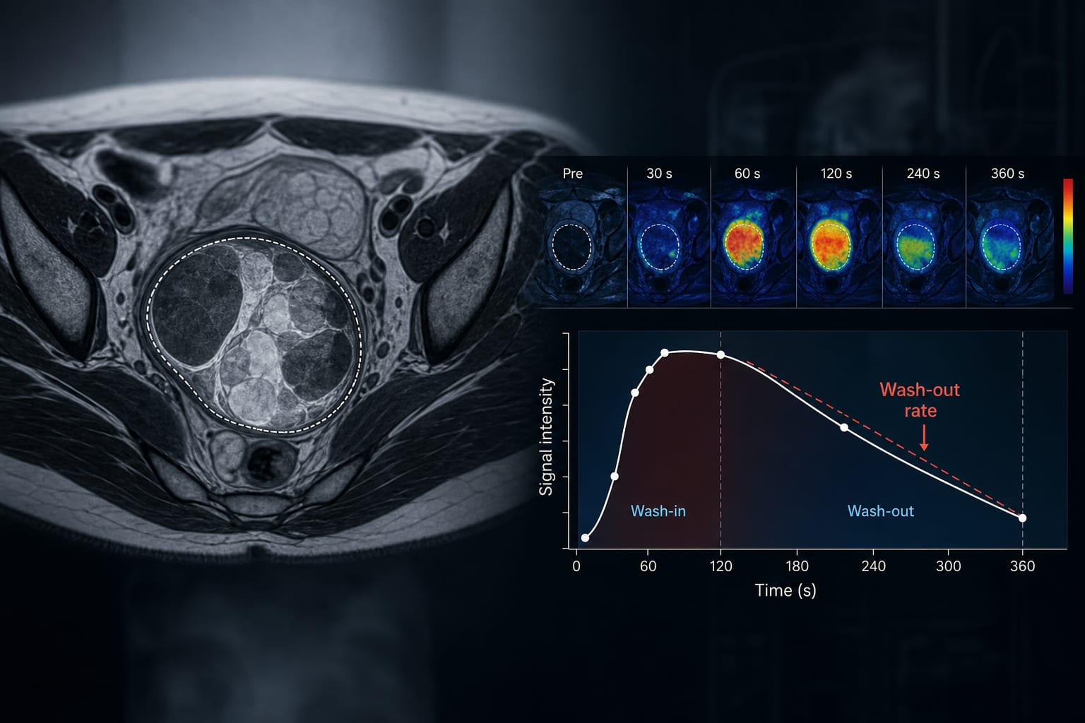
Abdominal CT revealed a heterogeneous mass with calcifications and fatty components measuring approximately 5 cm in greatest dimension, adherent to the anterior gastric wall.
Serum tumor markers were markedly elevated: AFP >60,500 ng/mL, NSE 63.8 ng/mL, and LDH 575 U/L, supporting a diagnosis of germ cell tumor.
Surgical exploration confirmed transmural growth of the tumor; complete resection was achieved with negative margins.
Histopathology identified a grade III immature teratoma, indicating malignant potential.
Postoperative recovery was uneventful with declining inflammatory markers and no immediate complications.
Clinical Implications
Clinicians should consider gastric teratoma in the differential diagnosis of neonatal abdominal masses, especially when imaging reveals calcifications and fatty components. Elevated serum AFP and other tumor markers can aid in diagnosis and monitoring. Complete surgical excision remains the cornerstone of treatment, and multidisciplinary collaboration is essential for timely diagnosis and management of these rare tumors.
Conclusion
This case underscores the rarity and diagnostic challenges of immature gastric teratomas in neonates, emphasizing the value of integrated imaging, tumor marker evaluation, and surgical management to achieve favorable outcomes.
References
Case Study Report 2024 -- Immature Gastric Teratoma in a Neonate