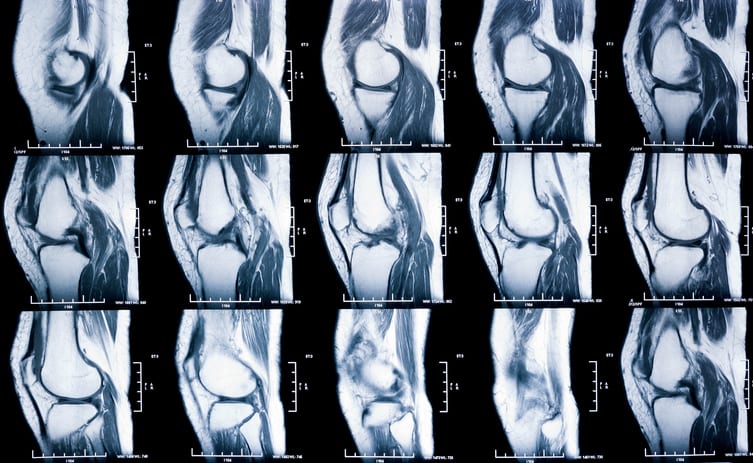
Assessment of Knee Injuries in Professional Soccer Players Using Semi-Quantitative MRI Scoring
Overview
This study evaluated knee joint health in 69 professional soccer players using semi-quantitative MRI scoring (WORMS) during their medical examinations. It found a high prevalence of prior knee injuries and variable days missed due to injury, with MRI providing objective assessment correlating with clinical history.
Background
Soccer is associated with repetitive and acute knee joint injuries, including articular cartilage lesions that may be symptomatic or asymptomatic. Such injuries can impair athletic performance and shorten careers. Medical examinations increasingly incorporate MRI to objectively assess joint health, with the Whole-Organ MRI Score (WORMS) being a widely used semiquantitative tool. This study aimed to test the reliability of WORMS in professional soccer players and its relationship to clinical data and injury-related missed playing time.
Mean days missed due to injury (prior injury group)
200 ± 232 (range 5–1105)
Total knee MRI scans analyzed
138 (both knees of 69 players)
Key Findings
69 male professional soccer players underwent bilateral 3.0-T knee MRI during medical examinations.
22 players (32%) had documented prior knee injuries, including ligament, tendon, meniscus, and cartilage lesions.
Mean days missed due to knee injuries was significantly higher in players with prior injuries (200 ± 232 days) compared to the overall cohort.
WORMS was adapted and used to semiquantitatively assess cartilage, bone marrow edema, osteophytes, synovitis, cysts, and ligament integrity.
Three experienced musculoskeletal radiologists independently scored MRI images blinded to clinical data, demonstrating reliability of the scoring method.
No players were excluded for severe knee problems at the time of medical examination, indicating subclinical or chronic changes were common.
Clinical Implications
Semi-quantitative MRI scoring using WORMS provides a reliable and objective method to detect and quantify knee joint abnormalities in professional soccer players, including those without overt symptoms. Identifying subclinical cartilage and ligament changes may help guide clinical management and injury prevention strategies. Incorporating MRI into routine medical examinations can improve assessment of player joint health and potentially reduce missed playing time.
Conclusion
Semi-quantitative MRI assessment during medical examinations is a valuable tool for detecting knee joint pathology in professional soccer players, correlating with prior injury history and missed playing days. This approach supports comprehensive evaluation to maintain player health and performance.
References
Study Authors/Medical Association of Hamburg/2020 -- Assessment of Knee Injuries in Professional Soccer Players Using Semi-Quantitative Magnetic Resonance Imaging Scoring
by Goetz Hannes Welsch, Anna-Maria Behr, Karl-Heinz Frosch, Enver Tahir, Milena Pachowsky, Frank Oliver Henes, Gerhard Adam, Kai-Jonathan Maas, Malte Lennart Warncke