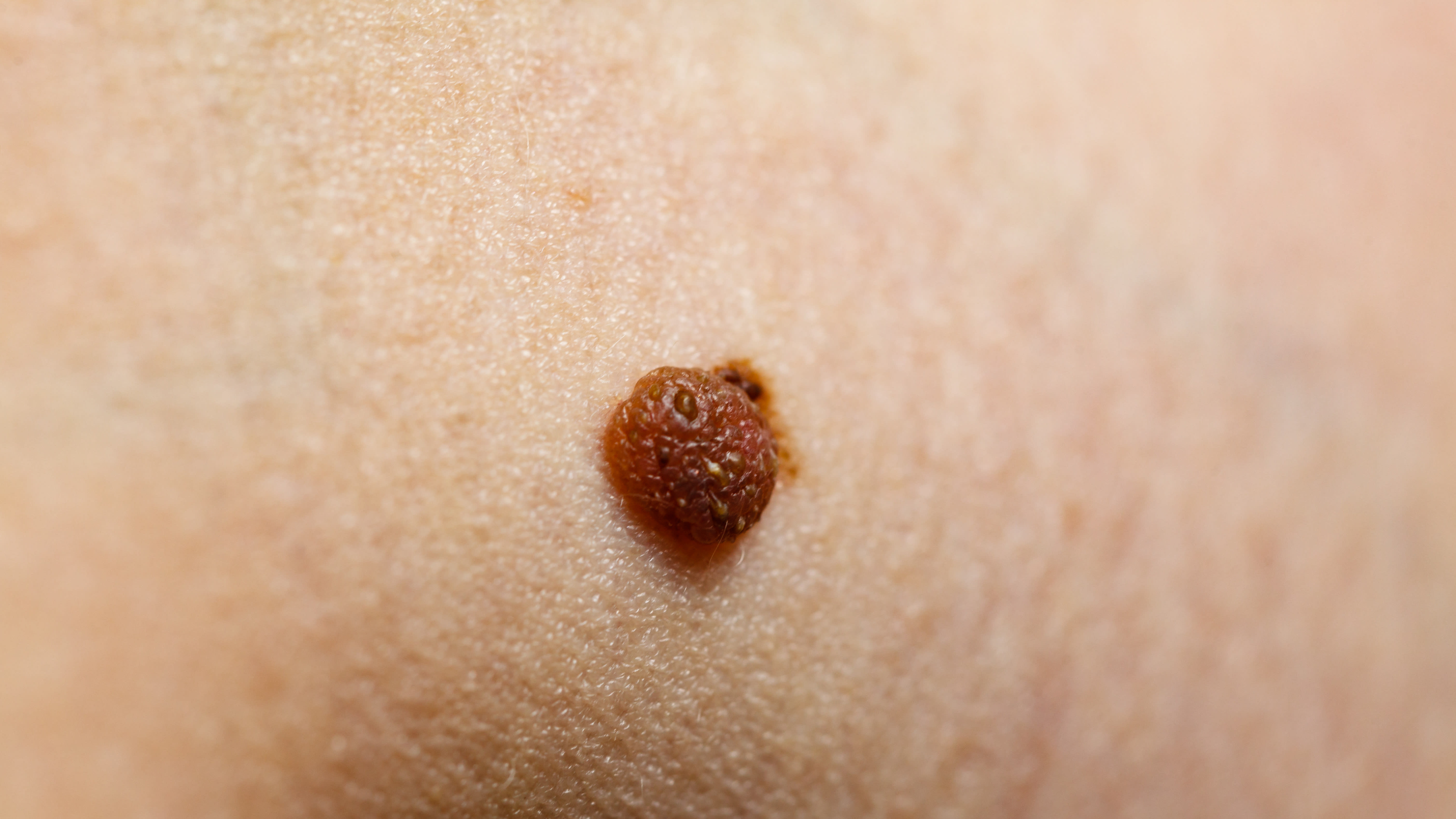
Deep Neural Network Framework for Automated Skin Lesion Segmentation
Overview
This study presents a robust framework combining traditional image enhancement and deep neural networks for automated segmentation of skin lesions in dermoscopic images. The approach addresses challenges such as lesion variability and artifacts, improving segmentation accuracy and reliability critical for skin cancer diagnosis.
Background
Skin cancer, including melanoma and non-melanoma types, represents a significant global health burden with millions of new cases annually. Early detection is vital, especially for melanoma due to its aggressive nature and high mortality. Dermoscopy enhances lesion visualization but presents segmentation challenges due to irregular lesion boundaries and artifacts. Conventional segmentation methods relying on manual features often fail to adapt to lesion diversity, necessitating advanced automated techniques.
Data Highlights
The proposed framework integrates morphological operations and Wiener filtering for pre-processing, followed by a deep neural network supported by Otsu’s thresholding for post-processing. This hybrid approach improves image quality and segmentation precision, addressing noise, contrast variability, and complex lesion characteristics inherent in dermoscopic images.
Key Findings
Skin cancer incidence is rising globally, with melanoma requiring early and accurate detection due to rapid metastasis.
Dermoscopy provides enhanced lesion visualization but complicates segmentation due to irregular boundaries and artifacts like hair and markings.
Traditional segmentation techniques relying on manual features are insufficient for complex dermoscopic images.
The proposed framework combines traditional image enhancement (morphological operations, Wiener filtering) with a deep neural network and Otsu’s thresholding for robust segmentation.
U-Net and its variants are effective but often computationally inefficient; the hybrid approach aims to balance accuracy and efficiency.
Advanced AI methods improve segmentation accuracy but face challenges in computational demand and generalization across diverse datasets.
Clinical Implications
Integrating advanced deep learning with traditional image enhancement techniques can significantly improve the accuracy and consistency of skin lesion segmentation in clinical practice. This facilitates more reliable computer-aided diagnosis, potentially leading to earlier detection and better management of skin cancers. Adoption of such robust frameworks may reduce diagnostic subjectivity and improve patient outcomes.
Conclusion
The hybrid framework leveraging both traditional image processing and deep neural networks offers a promising solution for the automated segmentation of skin lesions, addressing key challenges in dermoscopic image analysis. This approach enhances diagnostic precision, supporting improved clinical decision-making in skin cancer care.
References
American Cancer Society/National Cancer Institute/Global Skin Cancer Data -- Skin Cancer Statistics and Impact
Literature on U-Net and Advanced Segmentation Models -- Medical Image Segmentation Techniques
by Khlood M Mehdar, Toufique A Soomro, Ahmed Ali, Faisal Bin Ubaid, Muhammad Irfan, Hanan T Halawani, Aisha M Mashraqi, Sabah Elshafie Mohammed Elshafie, Abdullah A Asiri, Muawia Abdelkafi Magzoub