Clinical Report: Imaging Characteristics of Hepatic Parasitic Infections
Overview
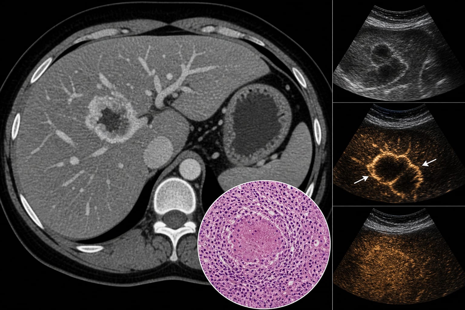
This case report highlights the diagnostic challenges posed by hepatic parasitic infections that can mimic malignancies on imaging modalities such as CT and ultrasound. A 38-year-old female was diagnosed with focal hepatic schistosomiasis after imaging suggested intrahepatic cholangiocarcinoma, emphasizing the importance of considering atypical infections in differential diagnoses.
Background
The differential diagnosis of focal liver lesions is a significant challenge in clinical radiology and oncology. Atypical presentations of chronic parasitic infections, such as schistosomiasis, can overlap with imaging features of malignancies, such as ill-defined borders and enhancement patterns, leading to potential misdiagnosis. Understanding these overlaps is crucial for accurate diagnosis and appropriate management.
Data Highlights
No numerical data or trial data available in the article.
Key Findings
A 38-year-old female presented with a left hepatic mass initially suspected to be intrahepatic cholangiocarcinoma based on imaging findings.
Normal serological tumor markers contradicted the initial imaging findings, raising suspicion for an atypical infection.
Contrast-enhanced ultrasound revealed atypical vascular patterns suggestive of hepatic paragonimiasis.
Histopathological analysis confirmed necrotizing granulomatous inflammation consistent with schistosomiasis.
The patient's history of residence in an endemic region for Schistosoma japonicum was pivotal in diagnosis.
Clinical Implications
Clinicians should consider atypical parasitic infections in the differential diagnosis of focal liver lesions, especially when imaging findings are discordant with serological results. Early recognition and accurate diagnosis, supported by serological testing, can prevent unnecessary invasive procedures and guide appropriate treatment.
Conclusion
This case underscores the importance of broadening the differential diagnosis to include atypical parasitic infections when faced with discordant imaging and serological findings. Accurate diagnosis, informed by patient history, is essential for effective management of hepatic lesions.