Management approaches, associated complications, and patient outcomes in spontaneous cerebellar hemorrhage: findings from a Swedish single-center observational study - Scorecard - MDSpire
Advertisement
Management approaches, associated complications, and patient outcomes in spontaneous cerebellar hemorrhage: findings from a Swedish single-center observational study
Clinical Scorecard: Management approaches, associated complications, and patient outcomes in spontaneous cerebellar hemorrhage: findings from a Swedish single-center observational study
At a Glance
Category
Detail
Condition
Spontaneous cerebellar hemorrhage (sCH), a subset of intraparenchymal brain bleedings
Key Mechanisms
Restricted posterior fossa volume leads to brainstem compression, obstructive hydrocephalus, and death from relatively small hematomas (>15 mL)
Target Population
Patients with spontaneous cerebellar hemorrhage, including elderly and those on antithrombotic therapy
Care Setting
Neurointensive care unit (NIC) at a tertiary referral center with neurosurgical intervention capabilities
Key Highlights
Surgical evacuation combined with external ventricular drainage (EVD) is standard for large sCH with brainstem compression or hydrocephalus.
Conservative management may suffice in awake patients with moderately large hematomas (~15 mL) without hydrocephalus or severe mass effect.
Optimal surgical strategies and NIC management targets remain poorly defined; institutional preference is suboccipital decompression with hematoma evacuation plus EVD.
Guideline-Based Recommendations
Diagnosis
Use CT imaging to assess hematoma size (>3 cm/15 mL), brainstem compression, and hydrocephalus.
Clinical assessment including Glasgow Coma Scale (GCS) to evaluate neurological status.
Management
Surgical intervention recommended for sCH with brainstem compression, clinical deterioration, acute hydrocephalus, or hematoma diameter >3 cm/15 mL.
EVD alone may be considered in patients with minor sCH but substantial intraventricular hemorrhage and acute hydrocephalus.
Conservative management may be appropriate for awake patients with moderately large hematomas without hydrocephalus or severe mass effect.
Monitoring & Follow-up
Neurointensive care with ICP/CPP monitoring and management extrapolated from traumatic brain injury and subarachnoid hemorrhage protocols.
Close neurological observation for clinical deterioration in conservatively managed patients.
Risks
Risk of upward herniation with EVD alone due to unresolved brainstem compression.
Potential exacerbation of hematoma growth and operative complications in patients on antithrombotic therapy.
Uncertainty regarding futility thresholds for surgery in elderly or frail patients.
Patient & Prescribing Data
194 patients with spontaneous cerebellar hemorrhage admitted to NIC unit over 2008–2024, including elderly and those on antithrombotic therapy
Surgical treatment favored in patients with neurological compromise and significant mass effect; conservative management viable in select awake patients; EVD alone reserved for minor sCH with hydrocephalus.
Clinical Best Practices
Perform suboccipital bone decompression with hematoma evacuation combined with EVD for large sCH with brainstem compression.
Consider duraplasty and posterior arch removal for additional decompression if posterior fossa remains constricted.
Select patients younger than 85 years with limited comorbidity and moderate-to-large hematomas for neurosurgical intervention.
Manage patients in NIC with focus on optimizing cerebral physiology using ICP/CPP protocols adapted from TBI and subarachnoid hemorrhage care.
Consult neurosurgery early for all intracranial hemorrhages and tailor treatment decisions based on clinical and radiological findings.
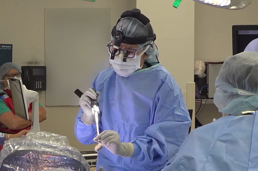
In this procedural case review, vascular surgeon Dr. Samuel Steerman and neurosurgeon Dr. Shannon Clark collaborate to perform an anterior lumbar interbody fusion (ALIF).