CT radiomics combined with clinical and radiological factors predict hematoma expansion in hypertensive intracerebral hemorrhage
-
By
-
Fei Yu
-
Mingguang Yang
-
Cheng He
-
Yanli Yang
-
Ying Peng
-
Hua Yang
-
Hong Lu
-
Heng Liu
-
July 11, 2024
-
Clinical Scorecard: Integrating CT Radiomics with Clinical and Imaging Factors to Forecast Hematoma Growth in Hypertensive Intracerebral Hemorrhage
At a Glance
| Category | Detail |
|---|
| Condition | Hypertensive intracerebral hemorrhage (HICH) with risk of hematoma expansion (HE) |
| Key Mechanisms | Hematoma expansion worsens patient outcomes; CT radiomics combined with clinical and imaging features improves HE prediction |
| Target Population | Patients with hypertensive intracerebral hemorrhage undergoing CT scans within 24 hours of onset |
| Care Setting | Emergency and neurology departments in hospital settings with CT imaging capabilities |
Key Highlights
- Hematoma expansion occurs in 13–38% of ICH patients and significantly increases risk of death or disability.
- Radiomics analysis of CT images combined with clinical and imaging data improves prediction accuracy of HE (AUC up to 0.960).
- A hybrid model integrating radiomics, clinical factors, and imaging signs provides an intuitive nomogram for clinical decision support.
Guideline-Based Recommendations
Diagnosis
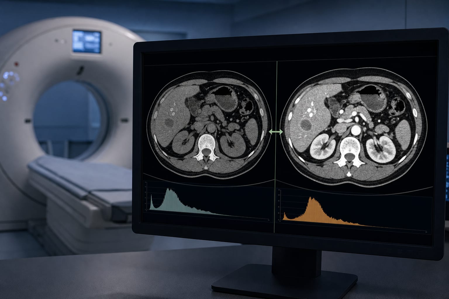
- Perform initial CT scan within 24 hours of symptom onset to assess hematoma volume and location.
- Use CT radiomics to extract quantitative imaging features for HE risk assessment.
- Combine clinical data (e.g., hypertension history, lab tests) and conventional imaging signs for comprehensive evaluation.
Management
- Identify patients at high risk of HE early to guide timely intervention aimed at reducing hematoma volume.
- Consider interventions that reduce hematoma size by 2–4 mL to decrease mortality or disability risk by 10–20% at 90 days.
Monitoring & Follow-up
- Repeat CT scan within 24 hours to measure hematoma volume changes and confirm HE.
- Use standardized hematoma volume measurement techniques (e.g., 3D Slicer software) for consistent monitoring.
Risks
- Exclude patients with secondary causes of ICH (trauma, vascular malformations, tumors) to avoid confounding HE prediction.
- Ensure high-quality CT images with clearly delineated hematoma borders for accurate radiomics analysis.
Patient & Prescribing Data
Patients with first hypertensive intracerebral hemorrhage and CT imaging within 24 hours
Early identification of HE risk via integrated radiomics and clinical models supports targeted interventions to improve outcomes
Clinical Best Practices
- Use a multidisciplinary approach involving neurologists and experienced neuroimaging physicians for data collection and interpretation.
- Apply multiple imputation methods to handle missing clinical or imaging data when below 20% missingness.
- Validate predictive models with independent test sets to ensure generalizability and reliability.
- Utilize nomograms derived from hybrid models to facilitate clinical decision-making and risk communication.
References