Optical Coherence Tomography Indicators of Posterior Scleritis Across Various Disease Activity Stages
By
Sameera Nayak
Niroj Kumar Sahoo
Udit Ajmani
P. Srinivas Rao
Raja Narayanan
Ritesh Narula
Rajeev R. Pappuru
July 10, 2026
Clinical Scorecard: Optical Coherence Tomography Indicators of Posterior Scleritis Across Various Disease Activity Stages
At a Glance
Category Detail
Condition Posterior Scleritis Key Mechanisms Inflammation of the sclera involving adjacent episclera in the posterior segment of the eye. Target Population Patients diagnosed with posterior scleritis. Care Setting Tertiary eye care center
Key Highlights
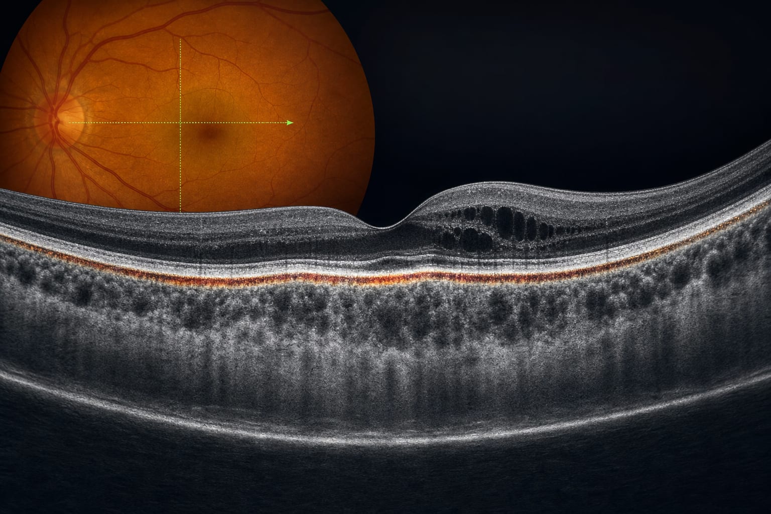
Acute PS demonstrated 100% choroidal thickening and 97.7% RPE–choroidal bump. Resolving PS showed 92.9% prevalence of hyperreflective dots in the inner and subretinal spaces. Resolved PS was characterized by 54.5% variable reflectivity of the ellipsoid zone. Guideline-Based Recommendations
Diagnosis
Diagnosis of PS is based on clinical presentation, ultrasonography, and fundus fluorescein angiography. Management
Treatment involves systemic corticosteroids initiated within 2 weeks of symptom onset. Monitoring & Follow-up
OCT characteristics can aid in identifying the stage of disease activity in PS. Risks
Posterior scleritis can be potentially sight-threatening. Patient & Prescribing Data
Patients with clinically diagnosed posterior scleritis.
Early diagnosis and treatment are crucial to prevent vision loss.
Clinical Best Practices
Utilize OCT for noninvasive analysis of the retina–choroidal complex. Systematically evaluate OCT images using predefined parameters. Related Resources & Content