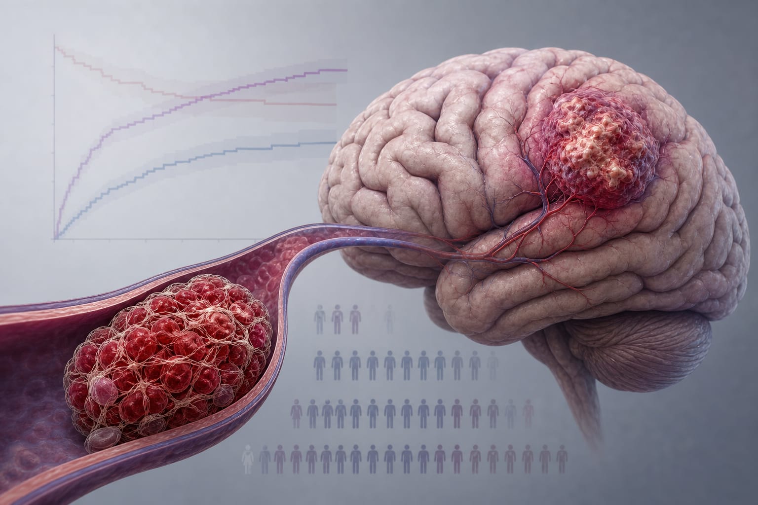
Clinical Scorecard: A Prospective Investigation of Keyhole Craniotomy and Stereotactic Brachytherapy for Newly Diagnosed Brain Oligometastases
At a Glance
Category
Detail
Condition
Metastatic brain tumors (MBTs) presenting as newly diagnosed brain oligometastases
Key Mechanisms
Combination of minimally invasive keyhole craniotomy (MIKC) for tumor resection and Cesium-131 (Cs-131) stereotactic brachytherapy (SBT) for targeted radiation delivery
Target Population
Adults with 1–6 newly diagnosed brain metastases, KPS ≥ 60, ECOG ≤ 2, requiring craniotomy and radiation therapy
Care Setting
Single-center prospective clinical setting with neurosurgical and radiation oncology collaboration
Key Highlights
Cs-131 SBT offers advantages over I-125 including shorter half-life (9.7 days), higher dose rate, better dose distribution, and reduced radiation necrosis.
MIKC reduces surgical trauma, accelerates recovery, and allows access to difficult tumor locations compared to conventional craniotomy.
Combined MIKC and Cs-131 SBT aims to improve local tumor control, reduce radiation necrosis, and enhance functional outcomes in brain oligometastases.
Guideline-Based Recommendations
Diagnosis
Use brain MRI with gadolinium to evaluate tumor location, size, and progression pre- and post-intervention.
Assess functional status with Karnofsky Performance Scale (KPS) and Eastern Cooperative Oncology Group (ECOG) scale.
Evaluate neurocognitive function using Mini-Mental State Examination (MMSE).
Assess quality of life with Functional Assessment of Cancer Therapy-Brain (FACT-Br) questionnaire.
Management
Perform surgical resection via minimally invasive keyhole craniotomy targeting dominant or largest metastasis when SRS is likely ineffective.
Implant Cs-131 stranded seeds into resection cavity for stereotactic brachytherapy with seed activity of 3–5 mCi each.
Calculate seed number based on tumor volume approximated as a sphere and resection cavity surface area.
Monitoring & Follow-up
Follow patients every two months with MRI and clinical assessments for up to two years or until local tumor progression.
Monitor for radiation necrosis, neurocognitive impairment, and neurological deficits.
Use Response Assessment in Neuro-Oncology (RANO)-Brain Metastasis criteria to assess local progression.
Risks
Radiation necrosis risk is reduced with Cs-131 compared to I-125 due to shorter half-life and improved radiobiologic properties.
Potential neurological complications and neurocognitive decline associated with whole brain radiotherapy (WBRT) are minimized with targeted SBT.
Patient & Prescribing Data
Adults newly diagnosed with 1–6 brain metastases, KPS ≥ 60, ECOG ≤ 2, excluding those with >6 lesions, prior radiosurgery or WBRT to same lesion, hemorrhage history, or life expectancy <3 months.
Cs-131 SBT combined with MIKC provides effective local control with fewer complications and improved functional outcomes; seed activity tailored per tumor volume and cavity size.
Clinical Best Practices
Select patients based on functional status and tumor characteristics to optimize benefit from surgery and brachytherapy.
Utilize image guidance and minimally invasive techniques to maximize safe tumor resection and preserve neurological function.
Implement systematic follow-up with standardized neuro-oncology criteria and functional assessments to monitor treatment efficacy and toxicity.
Consider Cs-131 SBT for its favorable radiobiologic profile and reduced radiation necrosis risk compared to traditional isotopes.