Efficacy and safety of cysto-ventricular catheter implantation for space-occupying cysts arising from glioma and brain metastasis: a retrospective study - Scorecard - MDSpire
Advertisement
Efficacy and safety of cysto-ventricular catheter implantation for space-occupying cysts arising from glioma and brain metastasis: a retrospective study
Clinical Scorecard: Assessment of the Safety and Effectiveness of Cysto-Ventricular Catheter Placement for Treating Space-Occupying Cysts Associated with Gliomas and Brain Metastases: A Retrospective Analysis
At a Glance
Category
Detail
Condition
Space-occupying intracranial cysts associated with gliomas and brain metastases
Key Mechanisms
Cysto-ventricular catheter (CVC) placement enables drainage of cyst fluid into the ventricle to reduce cyst volume and relieve mass effect
Target Population
Patients with symptomatic tumor-associated intracranial cysts from glioma or brain metastases without solid tumor progression
Care Setting
Neurosurgical tertiary care center with stereotactic frame-based imaging-guided surgery
Key Highlights
Space-occupying cysts occur in 4% of malignant glioma patients and up to 10–12% after interstitial brachytherapy for glioma
CVC placement is a minimally invasive stereotactic procedure that drains cyst fluid into the ventricular system
CVC is effective in reducing cyst volume and improving symptoms without requiring tumor-specific treatment if no solid tumor progression is present
Guideline-Based Recommendations
Diagnosis
Confirm tumor diagnosis via stereotactic biopsy or resection prior to CVC placement
Use MRI to identify space-occupying cysts causing compression and to exclude solid tumor progression
Define cyst progression as increasing cyst volume on serial MRI imaging
Management
Indicate CVC placement for symptomatic cysts with increasing size and no radiological tumor progression
Perform stereotactic frame-based imaging-guided surgery under general anesthesia
Insert vancomycin-impregnated ventricular catheter through planned trajectory into cyst and ventricle
Fix catheter on skull with titanium clip and confirm placement with postoperative CT
Monitoring & Follow-up
Perform preoperative, immediate postoperative, and follow-up MRI volumetry of cysts using T2-weighted images
Assess clinical symptoms including focal neurological deficits, seizures, and signs of elevated intracranial pressure
Evaluate cyst progression-free survival as sustained cyst volume reduction over 12 months
Risks
Potential complications related to stereotactic surgery and catheter placement
Risk of cyst recurrence depending on underlying tumor pathology fluid production
Need for secondary CSF diversion procedures if required
Patient & Prescribing Data
Patients with glioma or brain metastasis-associated intracranial cysts causing symptoms and cyst enlargement
CVC placement provides effective cyst volume reduction and symptom relief in patients without solid tumor progression, with individualized indication by interdisciplinary tumor board
Clinical Best Practices
Use interdisciplinary tumor board to define indications for CVC placement
Employ stereotactic frame-based imaging fusion for precise catheter trajectory planning
Utilize vancomycin-impregnated catheters to reduce infection risk
Perform quantitative volumetry of cysts pre- and postoperatively for objective assessment
Monitor patients clinically and radiographically for at least 12 months to assess cyst progression-free survival
These 10 states make it more practical for physicians to participate in hospital ownership by aligning statutory structure, corporate practice of medicine rules, and population trends.
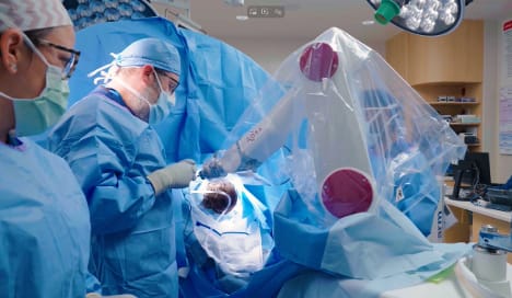
Phoenix Children’s continues to advance minimally invasive neurosurgical techniques with the integration of advanced robotics into Laser Interstitial Thermal Therapy (LITT) procedures