Clinical Scorecard: Density Assessment of Intracranial Arteriosclerosis via CT Imaging: Findings from a Population-Based Cohort Analysis
At a Glance
Category
Detail
Condition
Intracranial arteriosclerosis characterized by intracranial artery calcification (IAC)
Key Mechanisms
Calcification density as a marker of atherosclerotic disease stability; distinct morphological subtypes of IAC (medial and intimal) with differing determinants and cerebrovascular implications
Target Population
Older adults aged ≥ 55 years from the general population
Care Setting
Population-based observational cohort study with non-contrast-enhanced CT imaging
Key Highlights
IAC density may provide additional information beyond calcification volume, reflecting disease stability similar to coronary artery calcification.
Two morphological subtypes of IAC exist: medial (internal elastic lamina) and intimal (intimal layer), each with distinct cardiovascular risk factor associations and clinical outcomes.
Current literature on IAC density is limited, particularly in non-contrast CT imaging and its determinants remain unclear.
Guideline-Based Recommendations
Diagnosis
Use non-contrast-enhanced multidetector CT (MDCT) optimized for intracranial arteries to assess IAC presence, volume, and density.
Apply validated, reliable scoring methods for IAC assessment with high inter-rater reliability.
Management
Consider cardiovascular risk factors such as diabetes, hypertension, and smoking in the evaluation of IAC subtypes.
Recognize the potential differential impact of medial versus intimal calcifications on cerebrovascular outcomes.
Monitoring & Follow-up
Monitor IAC progression and density changes in longitudinal studies to understand their clinical implications.
Incorporate comprehensive cardiovascular and cerebrovascular risk profiling in follow-up assessments.
Risks
Medial calcifications are associated with diabetes, vascular diseases, white matter hyperintensities, dementia, and variable functional outcomes post-stroke therapies.
Intimal calcifications are linked to smoking, hypertension, and better collateral status on CT imaging.
Patient & Prescribing Data
Older adults aged ≥ 55 years undergoing CT imaging for intracranial arteriosclerosis assessment
No direct treatment data provided; understanding IAC density and subtype may inform risk stratification and therapeutic decisions in cerebrovascular disease.
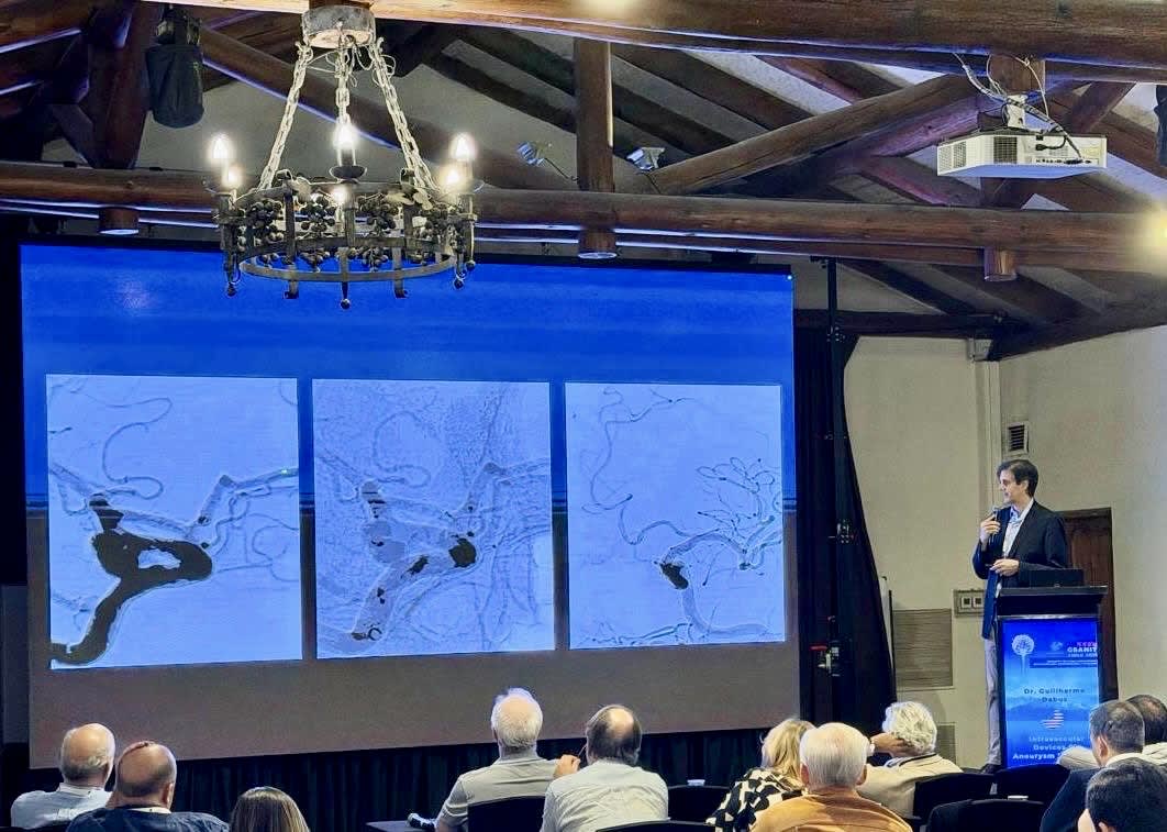
Guilherme Dabus, M.D., co-director of interventional neuroradiology at Baptist Health Miami Neuroscience Institute, served as a guest professor and invited speaker at the GSANIT (Grupo Sudamericano de Neurorradiología Intervencionista y Terapeutica) in Santa Cruz, Chile,