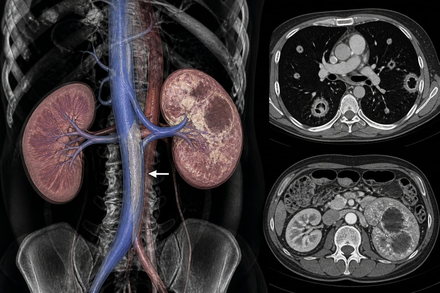
Klebsiella pneumoniae pyelonephritis and renal abscesses were associated with an infected thrombus extending from the renal vein into the inferior vena cava and cavitary septic pulmonary emboli
Target Population
A 47-year-old woman with untreated diabetes and persistent Klebsiella pneumoniae bacteremia
Care Setting
Acute hospital care involving antimicrobial therapy, anticoagulation, insulin treatment, imaging, and long-term follow-up
Key Highlights
The patient presented with dysuria, 11 days of fever, abdominal pain, and hypoxemia.
Blood and urine cultures grew Klebsiella pneumoniae, with bacteremia persisting despite meropenem.
CT showed pyelonephritis, renal abscesses, a near-occlusive renal vein–inferior vena cava thrombus, pulmonary arterial emboli, and bilateral peripheral cavitary nodules.
An 11-mm positive string test supported a hypermucoviscous phenotype, but hypervirulence was not genetically confirmed.
Ceftriaxone, levofloxacin, anticoagulation, and insulin treatment were followed by clearance of blood cultures by day 7.
By day 117, the thrombus and septic pulmonary emboli had nearly resolved.
Guideline-Based Recommendations
This single case report did not establish clinical guidelines or compare treatment strategies.
Diagnosis
Persistent Klebsiella pneumoniae bacteremia with peripheral cavitary pulmonary nodules should prompt evaluation for an occult septic venous source.
Venous imaging may be particularly relevant when urinary infection, renal abscesses, diabetes, or negative echocardiography is present.
Contrast-enhanced CT identified the renal infection, abscesses, venous thrombosis, and pulmonary abnormalities.
Transesophageal echocardiography showed no vegetation.
Management
Initial meropenem therapy was changed to ceftriaxone at meningitis dosing plus levofloxacin based on susceptibility and the need for exposure and penetration into deep abscesses.
Anticoagulation and insulin treatment were administered concurrently.
Nephrectomy, thrombectomy, and inferior vena cava filter placement were considered hazardous.
Continued medical management was selected because of the extensive infected thrombus and respiratory instability.
Monitoring & Follow-up
Blood cultures cleared by day 7.
CT findings worsened on day 10 despite subsequent clinical improvement.
Clinical, laboratory, and radiographic improvement by day 38 allowed intervention to be avoided.
Antimicrobials ended on day 49, and rivaroxaban was discontinued after near-complete resolution by day 117.
No recurrent thrombosis was observed on day 207.
Risks
Untreated diabetes may increase vulnerability to severe infection.
Renal vein–inferior vena cava septic thrombophlebitis may produce cavitary septic pulmonary emboli.
Early radiographic progression may occur despite later improvement.
A hypermucoviscous phenotype does not by itself establish genetic hypervirulence.
Patient & Prescribing Data
The patient was a 47-year-old woman with untreated diabetes and a hemoglobin A1c of 14.7%. The Klebsiella pneumoniae isolate was susceptible to ceftriaxone and levofloxacin.
Treatment included ceftriaxone, levofloxacin, anticoagulation, and insulin. More than 4 years after antimicrobial therapy ended, no infection, thrombosis, or septic pulmonary embolism had recurred.
Clinical Best Practices
Consider an occult septic venous source when Klebsiella pneumoniae bacteremia persists and peripheral cavitary nodules are present.
Use venous imaging to evaluate for septic thrombophlebitis when supported by the clinical context.
Interpret a positive string test as evidence of a hypermucoviscous phenotype rather than confirmation of hypervirulence.
Follow clinical, laboratory, microbiological, and radiographic findings during treatment.
Interpret the management outcome cautiously because it reflects a single case.
Related Resources & Content
Klebsiella pneumoniae Septic Pulmonary Embolism From Renal Vein Thrombosis — Nojo M, Ohta S, Fukuda Y, et al. International Journal of Infectious Diseases. 2026;171:109006. doi:10.1016/j.ijid.2026.109006.