Implementation of artificial intelligence in thoracic imaging—a what, how, and why guide from the European Society of Thoracic Imaging (ESTI) - Scorecard - MDSpire
Advertisement
Implementation of artificial intelligence in thoracic imaging—a what, how, and why guide from the European Society of Thoracic Imaging (ESTI)
Clinical Scorecard: A Comprehensive Guide to the Role of Artificial Intelligence in Thoracic Imaging: Insights from the European Society of Thoracic Imaging (ESTI)
At a Glance
Category
Detail
Condition
Thoracic imaging and related diagnostic processes
Key Mechanisms
Artificial intelligence (AI), including machine learning (ML) and deep learning (DL), applied to image analysis, lesion detection, segmentation, volumetry, and diagnostic support
Target Population
Patients undergoing thoracic imaging, including lung cancer screening and interstitial lung disease evaluation
Care Setting
Radiology departments and clinical settings utilizing thoracic imaging
Key Highlights
AI applications in thoracic imaging include noise reduction, lesion detection, segmentation, volumetry, and diagnostic support, but most lack sufficient clinical efficacy evidence.
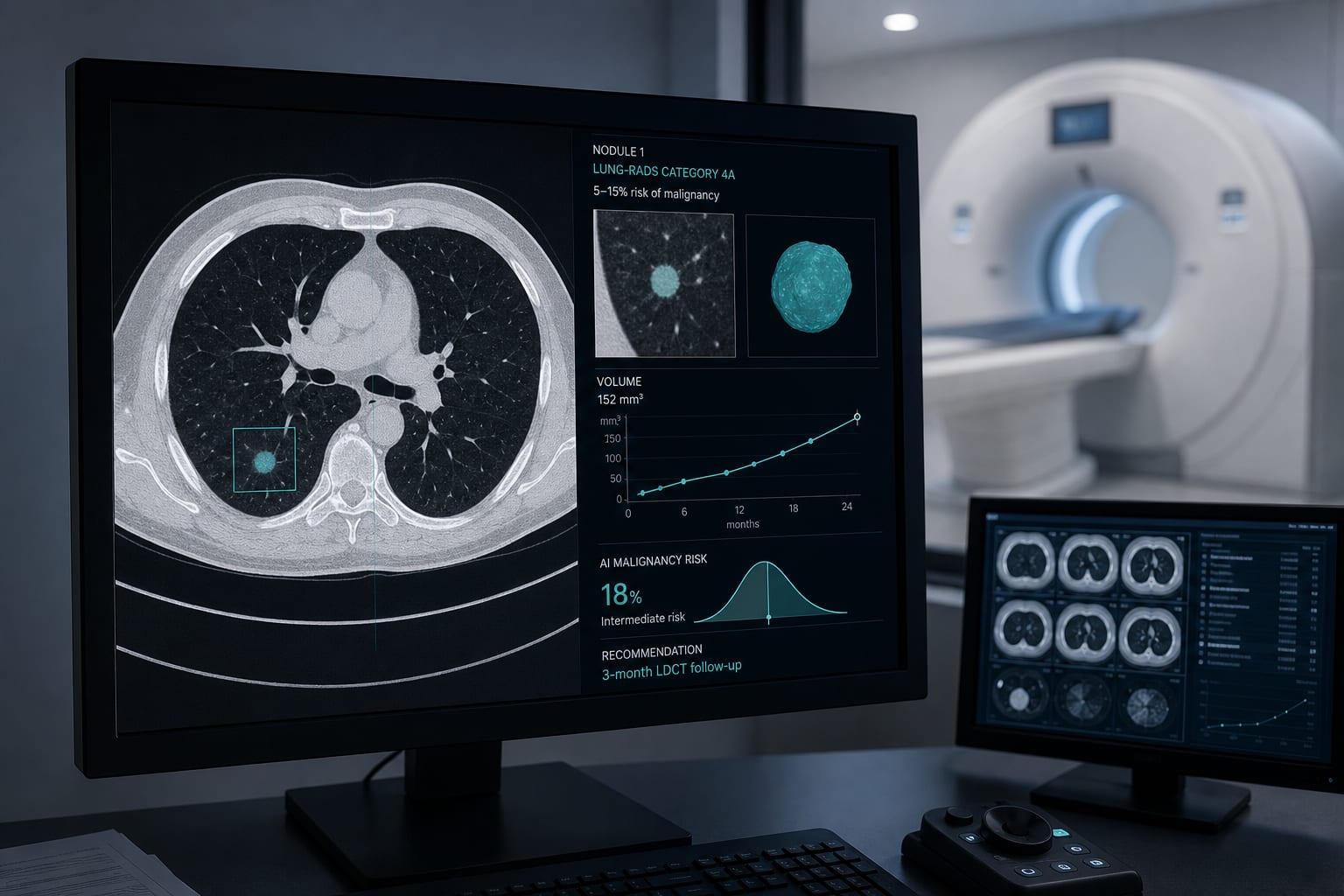
Computer-aided detection (CAD) tools enhanced by AI improve lung nodule detection sensitivity and reduce inter-observer variability, critical for lung cancer screening.
Clinical acceptance of AI tools depends on demonstrated utility, workflow efficiency, cost-effectiveness, and clear medicolegal accountability.
Guideline-Based Recommendations
Diagnosis
Use AI as an adjunct to radiologists for lesion detection and characterization, particularly in lung nodule detection.
Recognize that current AI models require rigorous validation due to methodological flaws and biases in training data.
Employ systematic approaches and adhere to published guidelines and checklists to ensure high-quality AI study design and evaluation.
Management
Integrate AI tools to augment radiologist workflow, aiming to reduce tedious tasks and improve diagnostic accuracy.
Consider hybrid radiology reports combining radiologist and AI-generated content with clear identification of AI contributions.
Implement AI solutions that demonstrate workflow speed improvements or diagnostic enhancements without excessive cost.
Monitoring & Follow-up
Maintain meticulous logs of AI decision history to ensure traceability and medicolegal accountability.
Use blockchain-based electronic medical records where possible to track AI algorithm versions and contributions over time.
Risks
Be aware of potential biases and systematic errors in AI models due to limited and non-representative training data.
Recognize the risk of false positives that may slow workflow or reduce efficiency.
Understand that lack of clinical validation may limit the usefulness of some AI applications.
Patient & Prescribing Data
Patients undergoing thoracic imaging, including those in lung cancer screening programs and with interstitial lung disease
AI tools improve detection sensitivity and reduce variability in lesion assessment but require careful integration to avoid workflow delays and ensure clinical utility.
Clinical Best Practices
Use AI to assist, not replace, radiologists in thoracic imaging interpretation.
Validate AI tools with multi-centric, well-curated datasets to minimize biases.
Adopt hybrid reporting systems that clearly delineate AI and human contributions.
Monitor AI performance continuously and document decision-making processes for accountability.
Prioritize AI solutions that enhance workflow efficiency and are cost-effective.
by Fergus Gleeson, Marie-Pierre Revel, Jürgen Biederer, Anna Rita Larici, Katharina Martini, Thomas Frauenfelder, Nicholas Screaton, Helmut Prosch, Annemiek Snoeckx, Nicola Sverzellati, Benoit Ghaye, Anagha P. Parkar