Clinical Scorecard: Temporary Flow Modulation Using Rapidly Resorbable Gelatin Microspheres in Yttrium-90 Radioembolization for Liver Cancer: A Preliminary Clinical Trial
At a Glance
Category
Detail
Condition
Primary or secondary liver cancer treated with Y-90 radioembolization
Key Mechanisms
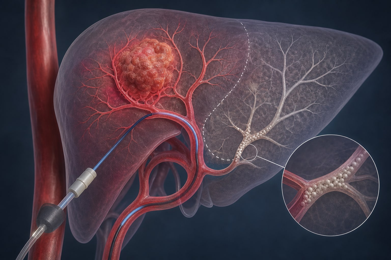
Rapid-resorbable gelatin microspheres temporarily embolize arteries supplying non-tumorous liver during Y-90 administration; the particles are intended to dissolve by approximately 70% within 24 hours and resorb completely within 3 days in vitro.
Target Population
The SEGWAY phase II, single-arm trial enrolled 20 adults with primary or secondary liver cancer, Child–Pugh class A, and non-tumorous liver volume of at least 50% of the Y-90 perfused liver volume.
Care Setting
Angiography-guided Y-90 radioembolization planning and treatment for selected patients with liver cancer.
Key Highlights
The prospective SEGWAY phase II, single-arm trial investigated whether rapidly resorbable gelatin microspheres protect non-tumorous liver by temporarily modulating arterial flow during Y-90 radioembolization.
The microspheres are physically crosslinked porcine gelatin and are designed to remain stable during delivery; in vitro, they are intended to dissolve by approximately 70% within 24 hours and resorb completely within 3 days.
Operators selected the arteries for protection, microsphere type, and final prescribed tumor absorbed dose; the protocol did not specify a target tumor absorbed dose or non-tumorous liver dose in advance.
The supplied excerpt describes the trial rationale and methods; it does not include clinical outcomes or quantitative dosimetric results.
Guideline-Based Recommendations
Diagnosis
Trial eligibility required primary or secondary liver cancer diagnosed by Gd-EOB-DTPA-enhanced MRI or biopsy.
Management
Planning included angiography and technetium-99m MAA imaging with planar imaging and SPECT/CT.
The study used partition-model dosimetry. Operators selected the final prescribed tumor absorbed dose and arteries receiving RGM; the protocol did not specify target tumor or non-tumorous liver doses in advance.
RGMs were selectively delivered through a 2.4-Fr microcatheter to arteries designated for protection from Y-90 microsphere delivery.
Monitoring & Follow-up
Planning included segmentation of the whole liver, tumor, and non-tumorous liver, followed by manual rigid registration of SPECT/CT images.
The estimated lung dose was below 30 Gy in the described planning approach.
Risks
Permanent embolic agents can prevent later transarterial treatment of the protected territory and may compromise regional liver function through persistent hypoxia.
Conventional gelatin sponge particles may take weeks to months to resorb, potentially causing persistent injury to treated arteries and corresponding liver parenchyma.
Vascular invasion and biliary–enteric anastomosis were exclusion criteria because either condition may confound image-based evaluation of post-radioembolization non-tumorous liver function.
Patient & Prescribing Data
SEGWAY eligibility included adults with primary or secondary liver cancer, Child–Pugh class A, no locoregional treatment of the same hepatic lobe in the preceding year, and non-tumorous liver volume at least 50% of the Y-90 perfused liver volume. Patients with vascular invasion or biliary–enteric anastomosis were excluded.
Y-90 radioembolization used glass or resin microspheres. Operators selected microsphere type, activity per microsphere, prescribed tumor absorbed dose, and arteries for RGM protection. The protocol encouraged a tumoricidal dose but specified no target tumor absorbed dose or non-tumorous liver dose.
Clinical Best Practices
Trial planning used angiography and technetium-99m MAA planar imaging and SPECT/CT.
The study used partition-model dosimetry based on segmented whole-liver, tumor, and non-tumorous liver volumes.
The excerpt states that operators could account for the RGM-protected territory in dosimetry planning; it does not provide enough detail to support a general best-practice recommendation.
Remove the claim that untreated liver volume was required to exceed 30% of overall non-tumorous liver volume unless supported by the full source.
Harold Burstein, MD, PhD, and Jennifer A. Ligibel, MD discuss new research from the Breast Cancer Weight Loss (BWEL) trial, as well as recommendations for lifestyle, diet and GLP1 agonists.
SOTIO Biotech’s CSO explains why drug developers should test their weakest assumptions early, how she builds innovative teams, and where ADCs could go next
Harold Burstein, MD, PhD, and Ana C. Garrido-Castro, MD discuss new results from the PANKU-Breast02 Study on a new dual-antibody drug conjugate, called izalontamab brengitecan (iza-bren), for metastatic triple-negative breast cancer.