Standardizing the estimation of ischemic regions can harmonize CT perfusion stroke imaging
By
Daan Peerlings
Edwin Bennink
Jan W. Dankbaar
Birgitta K. Velthuis
Bart J. Emmer
Jan W. Hoving
Charles B. L. M. Majoie
Henk A. Marquering
Henk van Voorst
Hugo W. A. M. de Jong
August 12, 2023
Clinical Scorecard: Harmonizing CT Perfusion Imaging for Stroke: A Standardized Approach to Estimating Ischemic Areas
At a Glance
Category Detail
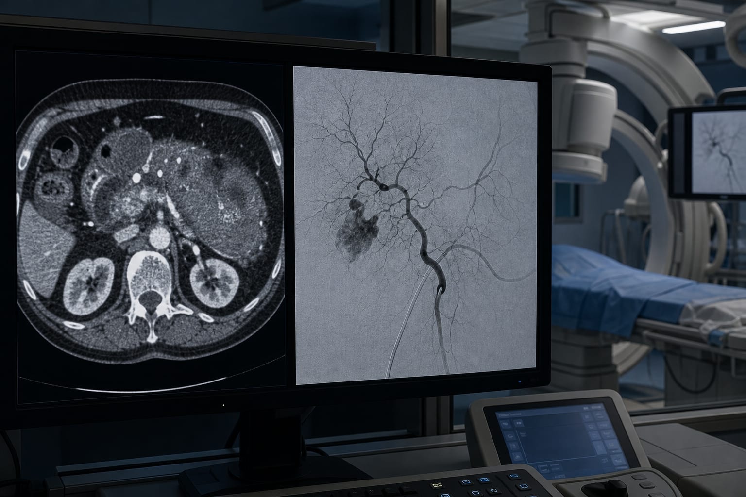
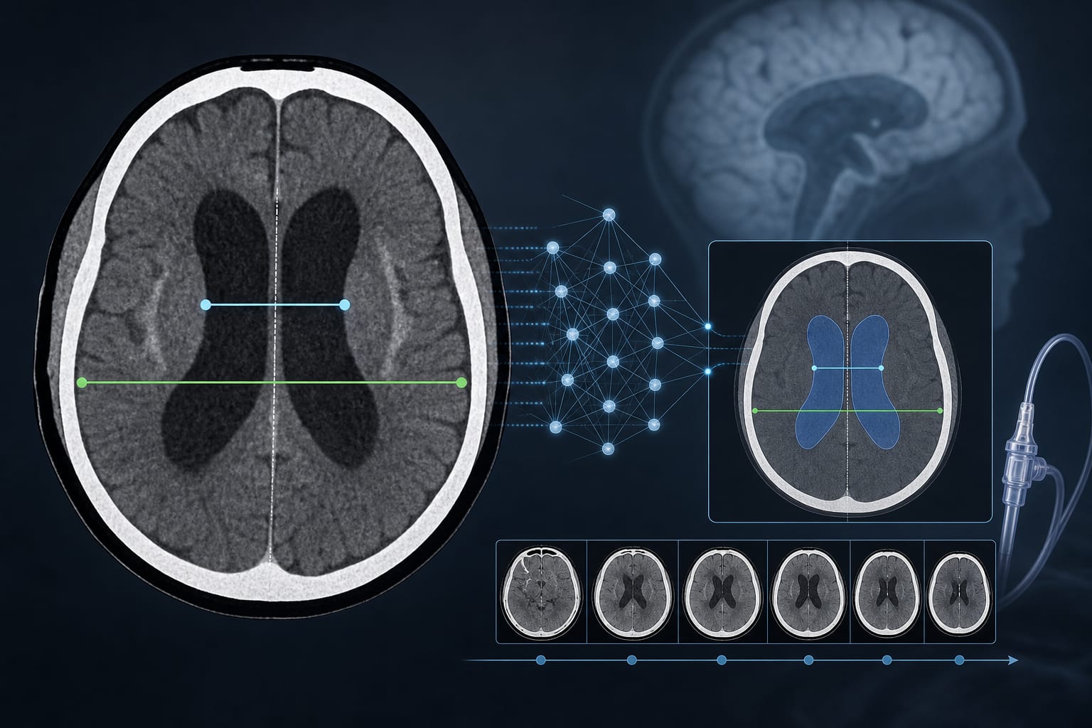
Condition Acute ischemic stroke Key Mechanisms CT perfusion imaging variability due to differences in scan protocols and vendor software affecting ischemic area estimation Target Population Patients with acute ischemic stroke undergoing CT perfusion imaging Care Setting Multicenter stroke centers performing CT perfusion imaging
Key Highlights
Multicenter CT perfusion imaging protocols vary substantially, impacting consistency of ischemic stroke evaluation. Scan acquisition parameters (tube voltage, exposure, timing) and vendor-specific software algorithms influence perfusion results. Standardization of CT perfusion protocols and vendor software may harmonize ischemic area estimation across centers. Guideline-Based Recommendations
Diagnosis
Use CT perfusion imaging with standardized acquisition parameters to improve ischemic stroke assessment consistency. Apply vendor-specific default thresholds cautiously due to variability in ischemic region estimation. Management
Consider harmonizing CT perfusion imaging protocols across centers to support reliable clinical decision-making. Incorporate vendor software characteristics when interpreting perfusion maps for treatment planning. Monitoring & Follow-up
Monitor variability in CT perfusion results related to scan protocol differences and software processing. Use phantom-based evaluations to assess and adjust for noise and protocol effects on perfusion imaging. Risks
Variability in imaging protocols and software may lead to inconsistent ischemic core and penumbra volume estimation. Inaccurate ischemic area estimation could affect treatment eligibility and outcomes. Patient & Prescribing Data
Acute ischemic stroke patients undergoing CT perfusion imaging in multicenter settings
Standardized imaging protocols and software harmonization are critical to ensure accurate ischemic area delineation guiding reperfusion therapies.
Clinical Best Practices
Request and document scan protocols (tube voltage, exposure, timing) for CT perfusion imaging at each center. Use anthropomorphic digital phantoms to simulate and evaluate center-specific scan protocols and software performance. Adhere to default vendor software settings but validate ischemic region thresholds against standardized references. Upsample or rescale perfusion maps appropriately to maintain spatial resolution consistency across software. Collaborate across centers to develop and implement standardized CT perfusion imaging protocols. References