Clinical Scorecard: Alterations in White Matter Associated with Alzheimer's Disease: Are They a Cause, Effect, or Coexisting Condition?
At a Glance
Category
Detail
Condition
Alzheimer's disease (AD)
Key Mechanisms
Tau-mediated axonal degeneration, amyloid-related cerebrovascular injury, and disrupted clearance of pathological proteins through perivascular pathways
Target Population
Individuals across the AD spectrum from cognitively unimpaired to dementia
Care Setting
Neurology and memory clinics with neuroimaging capabilities
Key Highlights
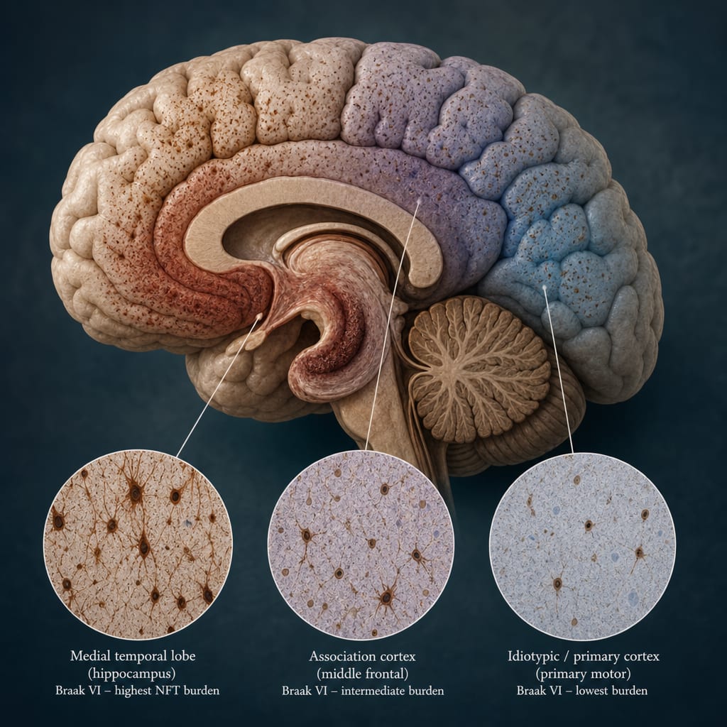
Cortical tau deposition precedes and parallels macroscopic white matter degeneration in AD.
White matter changes are likely a downstream consequence of AD pathology rather than an independent co-pathology.
Early tau-mediated white matter damage may occur before clinical symptoms, contributing to early cognitive decline.
Guideline-Based Recommendations
Diagnosis
Utilize tau-PET imaging to detect cortical tau deposition as a marker preceding white matter degeneration.
Incorporate MRI to assess white matter volume loss, focusing on limbic tracts and temporal-parietal regions for AD-related changes.
Consider spatial distribution of white matter abnormalities to differentiate AD pathology from cerebral small vessel disease.
Management
Recognize white matter pathology as a potential therapeutic target in AD progression.
Address mixed pathologies in clinical settings, especially coexisting AD and cerebrovascular disease.
Monitoring & Follow-up
Longitudinal imaging with tau-PET and MRI to track tau accumulation and white matter volume changes over time.
Monitor cognitive symptoms in relation to imaging findings to assess disease progression.
Risks
Potential misinterpretation of white matter changes due to overlapping cerebrovascular disease.
Limitations in current imaging sensitivity may obscure early microscopic white matter pathology.
Patient & Prescribing Data
AD spectrum patients including asymptomatic individuals with tau deposition
Early detection of tau-mediated white matter damage may inform timing of interventions before clinical symptom onset
Clinical Best Practices
Control for cerebrovascular confounders when interpreting white matter changes on MRI.
Use combined amyloid- and tau-PET imaging to clarify the sequence and contribution of pathologies.
Consider individual variability in white matter connectivity when assessing imaging data.
Recognize the importance of early tau pathology in white matter as a contributor to cognitive decline.