Clinical Scorecard: Automating C-arm Alignment for Standard Imaging Views in Orthopedic Procedures
At a Glance
Category
Detail
Condition
Challenges in obtaining correct standard fluoroscopic projections during orthopedic and trauma surgery
Key Mechanisms
Deep learning-based regression model predicting C-arm pose adjustments from 2D fluoroscopic images without additional hardware or preoperative CT
Target Population
Patients undergoing orthopedic or trauma surgery requiring fluoroscopic imaging
Care Setting
Intraoperative imaging in orthopedic and trauma surgical suites
Key Highlights
Incorrect C-arm positioning leads to poor standard projections, increasing risk of overlooked errors such as fracture malunion.
Current manual C-arm positioning relies on trial-and-error fluoroscopy, causing increased radiation exposure and time consumption.
Proposed CNN regression model predicts 5 degrees of freedom pose updates directly from 2D images, trained on simulated data without need for preoperative CT or external tracking.
Guideline-Based Recommendations
Diagnosis
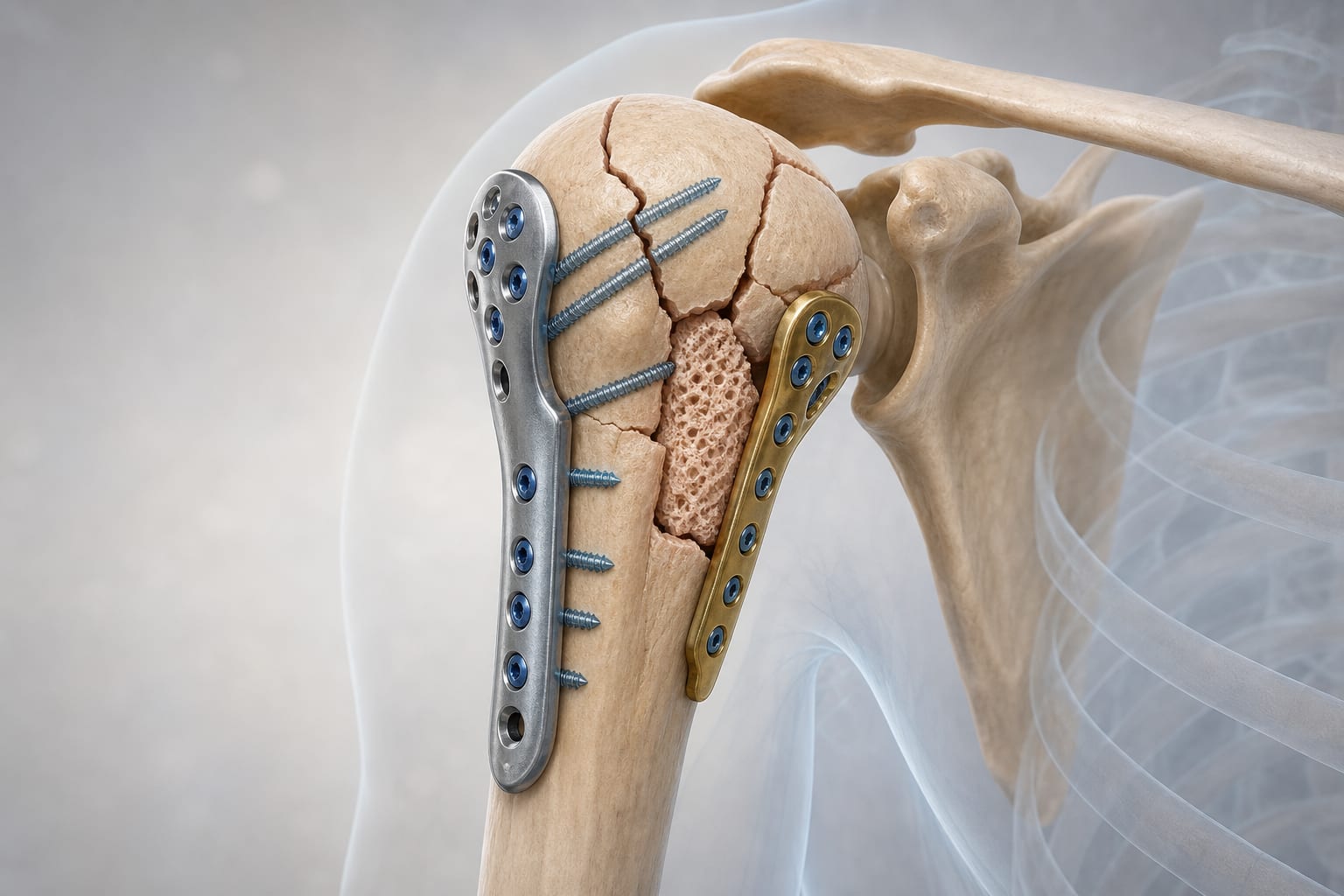
Use fluoroscopic imaging to verify fracture reduction and implant placement.
Recognize that standard projections are essential for accurate assessment but are often surgeon-dependent and variable.
Management
Employ automated C-arm positioning assistance systems that do not require external tracking hardware or preoperative CT scans.
Utilize deep learning models trained on simulated fluoroscopic images to guide C-arm alignment toward anatomy-specific standard views.
Monitoring & Follow-up
Continuously assess fluoroscopic image quality to ensure correct standard projections are achieved.
Monitor radiation exposure to patients and personnel, minimizing repeated imaging through improved positioning accuracy.
Risks
Incorrect projections can cause overlays and ambiguities, leading to missed errors such as malunion or implant misplacement.
Trial-and-error fluoroscopy increases radiation exposure and procedure time.
Dependence on external hardware or preoperative imaging may limit clinical applicability and workflow integration.
Patient & Prescribing Data
Orthopedic and trauma surgery patients requiring intraoperative fluoroscopic imaging
Automated C-arm positioning can reduce radiation exposure and improve accuracy of standard imaging views, potentially decreasing complications related to misaligned imaging.
Clinical Best Practices
Train operators in the use of deep learning-assisted C-arm positioning tools to reduce trial-and-error fluoroscopy.
Incorporate simulated training datasets to improve model accuracy and generalizability to different anatomies such as proximal femur and spine.
Avoid reliance on external tracking hardware or preoperative CT scans to facilitate broader clinical adoption.
Validate automated positioning approaches on real cadaveric or clinical fluoroscopic images before routine use.