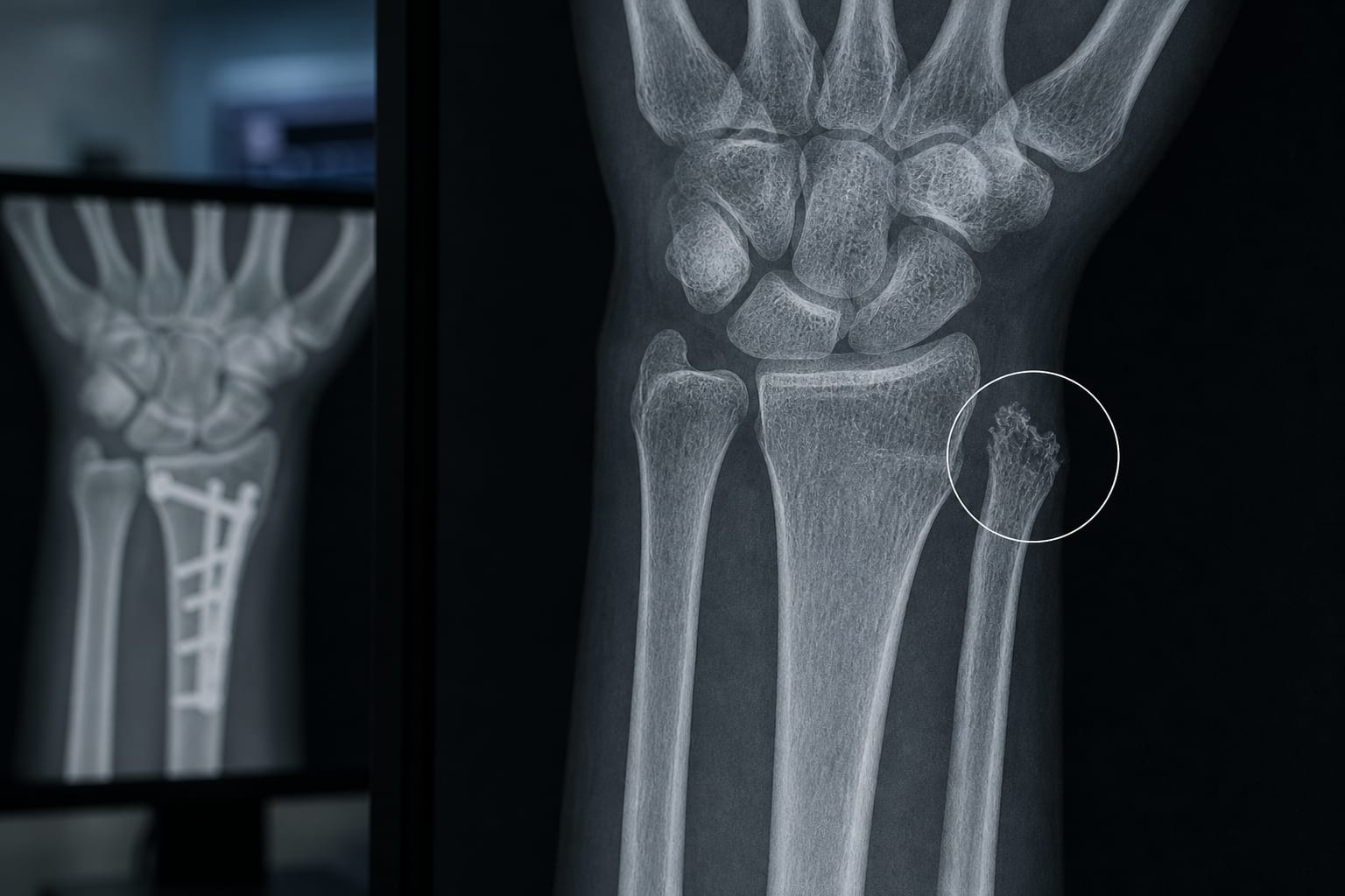
Ulnar Styloid Osteolysis: A Clinically Meaningful Finding Beyond Imaging?
-
By
-
Jan-Peter Grunz
-
Malek Othmani
-
Andreas Steven Kunz
-
November 9, 2026
Clinical Scorecard: Ulnar Styloid Osteolysis After Distal Radius Fracture: A Post-Traumatic Imaging Finding
At a Glance
| Category | Detail |
|---|---|
| Condition | Ulnar styloid osteolysis: delayed cortical resorption of the ulnar styloid without a visible ulnar fracture, reported after distal radius fractures. |
| Key Mechanisms | The biological basis is uncertain. Proposed explanations include true cortical bone loss, insertional remodeling, occult microfractures, or combinations of these; links to fracture severity, DRUJ biomechanics, and TFCC injury remain unconfirmed. |
| Target Population | People with distal radius fractures undergoing radiographic follow-up. |
| Care Setting | Musculoskeletal imaging and orthopedic follow-up after distal radius fracture. |
Key Highlights
- A retrospective study of 143 people with distal radius fractures found ulnar styloid osteolysis in 16.8%.
- Most lesions became apparent during the initial months after injury.
- Osteolysis was associated with comminution, dorsal angulation, diastasis, impaction, and active smoking.
- The clinical significance and biological substrate of the finding remain uncertain.
- Delayed erosive-appearing abnormalities may be misinterpreted as inflammatory or erosive arthropathies if their post-traumatic context is not recognized.
Guideline-Based Recommendations
Diagnosis
- The article describes delayed ulnar styloid cortical defects on follow-up radiographs after distal radius fracture, without a visible ulnar fracture.
- The study diagnosed the finding primarily using follow-up radiographs; the structural nature of the defects remains uncertain.
Management
- The source does not provide treatment recommendations for ulnar styloid osteolysis.
- The article identifies CT and MRI as potential modalities for future characterization; it does not establish a routine imaging pathway.
Monitoring & Follow-up
- Most lesions became apparent during the initial months following injury.
- The article calls for research into lesion morphology and temporal evolution using advanced imaging.
Risks
- Delayed erosive-appearing abnormalities may be misinterpreted as inflammatory or erosive arthropathies if their post-traumatic context is not recognized.
- Associations with DRUJ instability, TFCC injury, and post-traumatic wrist dysfunction are not established.
Patient & Prescribing Data
Retrospective analysis of 143 individuals with distal radius fractures; ulnar styloid osteolysis was identified in 16.8%.
No medication or treatment data are reported. The study describes an imaging finding and associated factors, not a treatment intervention.
Clinical Best Practices
- The article describes interpreting delayed ulnar styloid cortical defects in the context of prior distal radius fracture and follow-up imaging.
- Do not infer DRUJ dysfunction or TFCC injury from osteolysis alone; the article states that these relationships remain uncertain.
- The article cites expert consensus identifying CT as the most accurate technique for evaluating DRUJ instability and MRI as the preferred method for assessing TFCC pathology; these statements are not specific recommendations for osteolysis.
Related Resources & Content
- Freire et al. Contemporary systematic assessment of ulnar styloid osteolysis after distal radius fractures (as described in the source article)
- Expert consensus on imaging evaluation of DRUJ instability and TFCC pathology (as cited in the source article)
Based on findings from:
Ulnar styloid osteolysis: More than a radiographic curiosity?
Jan-Peter Grunz, Malek Othmani, Andreas Steven Kunz. European Radiology, 2026.
https://link.springer.com/article/10.1007/s00330-026-12819-7
This content is an AI-generated, fully rewritten summary based on a published scholarly article. It does not reproduce the original text and is not a substitute for the original publication. Readers are encouraged to consult the source for full context, data, and methodology.