Ultra-widefield optical coherence tomography angiography in diabetic retinopathy: from retinal lesions to choroidal metrics
By
Tianhong Luo
Yingshi Zou
Yali Gao
June 22, 2026
Clinical Scorecard: Comprehensive Analysis of Diabetic Retinopathy Using Ultra-widefield Optical Coherence Tomography Angiography: Insights into Retinal Lesions and Choroidal Metrics
At a Glance
Category Detail
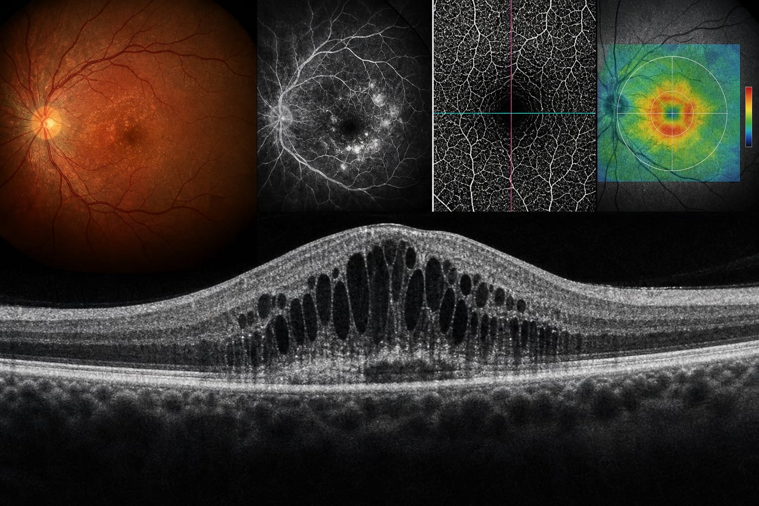
Condition Diabetic Retinopathy Key Mechanisms Ultra-widefield optical coherence tomography angiography (UWF-OCTA) provides depth-resolved vascular visualization and identifies key retinal microvascular lesions. Target Population Individuals with diabetes mellitus, particularly those at risk for visual impairment. Care Setting Clinical assessment and management of retinal vascular disorders.
Key Highlights
UWF-OCTA detects neovascularization (NV) with high sensitivity and specificity. Offers advantages over fluorescein angiography (FA) including non-invasiveness and depth-resolved imaging. Identifies peripheral non-perfusion areas (NPAs) and other retinal lesions that are often missed by conventional methods. Facilitates quantitative image analysis of retinal vascular parameters. Represents a promising tool for DR diagnosis and monitoring. Guideline-Based Recommendations
Diagnosis
Utilize UWF-OCTA for comprehensive assessment of diabetic retinopathy. Management
Consider UWF-OCTA as a complement to conventional angiography in clinical practice. Monitoring & Follow-up
Employ UWF-OCTA for ongoing monitoring of retinal changes in diabetic patients. Risks
Acknowledge limitations such as segmentation errors and smaller field of view compared to FA. Patient & Prescribing Data
Patients with diabetes mellitus at risk for diabetic retinopathy.
UWF-OCTA may enhance the detection and management of retinal complications associated with diabetes.
Clinical Best Practices
Incorporate UWF-OCTA into routine screening protocols for diabetic retinopathy. Educate patients about the benefits and limitations of UWF-OCTA compared to traditional imaging methods. Related Resources & Content