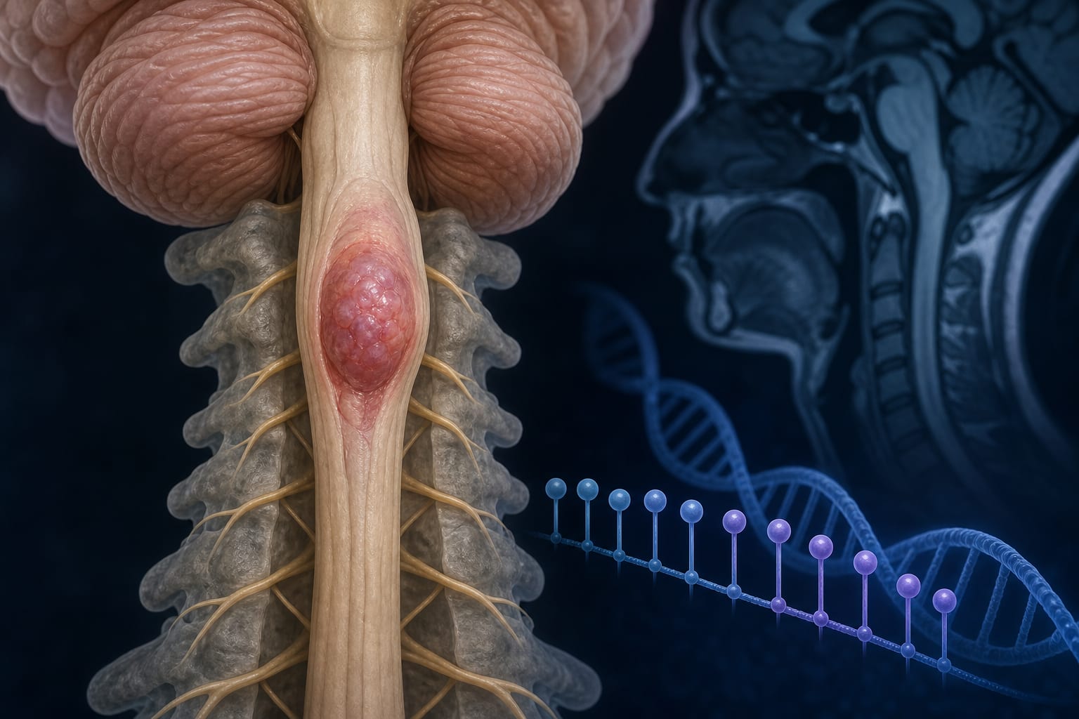
Clinical Scorecard: Analysis of Radiotherapy and Chemotherapy's Impact on Cerebral Volume in Oncology Patients: A Study on Brain Tumors
At a Glance
Category
Detail
Condition
Primary and metastatic brain tumors including gliomas, meningiomas, and metastatic lesions
Key Mechanisms
Uncontrolled proliferation of abnormal brain cells; tumor growth causing cerebral edema and neurological deficits; treatment effects from radiotherapy and chemotherapy on brain volume
Target Population
Adult patients aged 18–90 years diagnosed with intracranial masses undergoing radiotherapy and/or chemotherapy
Care Setting
Medical oncology departments with multidisciplinary management including surgical, radiotherapy, and chemotherapy interventions
Key Highlights
Brain tumors cause neurological and cognitive symptoms depending on tumor type, size, and location.
Radiotherapy and chemotherapy are critical adjuvant treatments, especially for aggressive tumors like glioblastoma multiforme.
Volumetric MRI assessments using tools like VolBrain and MRICloud enable objective measurement of brain and tumor volumes pre- and post-treatment.
Guideline-Based Recommendations
Diagnosis
Use conventional MRI sequences for anatomical tumor evaluation and diagnosis.
Perform volumetric measurements of brain structures and tumor volumes using validated software tools.
Histological classification per WHO criteria is essential for treatment planning and prognosis.
Management
Surgical resection is attempted when feasible to remove tumors.
Adjuvant radiotherapy with median doses around 60 Gy in 30–33 fractions is standard.
Chemotherapy is combined with radiotherapy for aggressive tumors to enhance treatment efficacy.
Follow-up MRI every 3 to 6 months for the first five years, then annually.
Monitoring & Follow-up
Regular MRI imaging to assess tumor response and cerebral volume changes.
Use of volumetric tools to objectively track brain structure changes during and after treatment.
Risks
Exposure to ionizing radiation is a known environmental risk factor for brain tumors.
Radiotherapy and chemotherapy may impact cerebral volume and brain structures, necessitating careful dose planning.
Patient & Prescribing Data
47 patients (11 females, 36 males) with primary or metastatic brain tumors treated with radiotherapy and/or chemotherapy
Median radiotherapy dose was 60 Gy over 30–33 fractions; 19 patients received whole brain irradiation at 40 Gy; treatment planning involved 3D dose distribution calculations.
Clinical Best Practices
Adopt a multidisciplinary approach integrating surgery, radiotherapy, and chemotherapy for brain tumor management.
Utilize advanced imaging and volumetric analysis tools for individualized treatment planning and monitoring.
Ensure ethical approval and retrospective data review for clinical studies involving imaging and treatment outcomes.
Tailor follow-up imaging schedules to tumor type and treatment response to optimize patient care.