Application of three-dimensional visualization technology on one-step PTCSL for complex hepatolithiasis
-
By
-
Yawen Cao
-
Qing Wu
-
Li-Yun Huang
-
Shunqian Wen
-
Zhi-Hao Lai
-
Shuai-Mei Luo
-
Zhao-Wei Ding
-
Cheng Li
-
Yong-Qing Ye
-
July 2, 2026
-
Clinical Scorecard: Utilization of 3D Visualization Techniques in One-Step PTCSL for Treating Complex Hepatolithiasis
At a Glance
| Category | Detail |
|---|
| Condition | Complex Hepatolithiasis |
| Key Mechanisms | Integration of 3D visualization technology into one-step PTCSL to enhance procedural precision and reduce intraoperative challenges. |
| Target Population | Patients with primary hepatolithiasis undergoing surgery. |
| Care Setting | Hepatobiliary surgery department |
Key Highlights
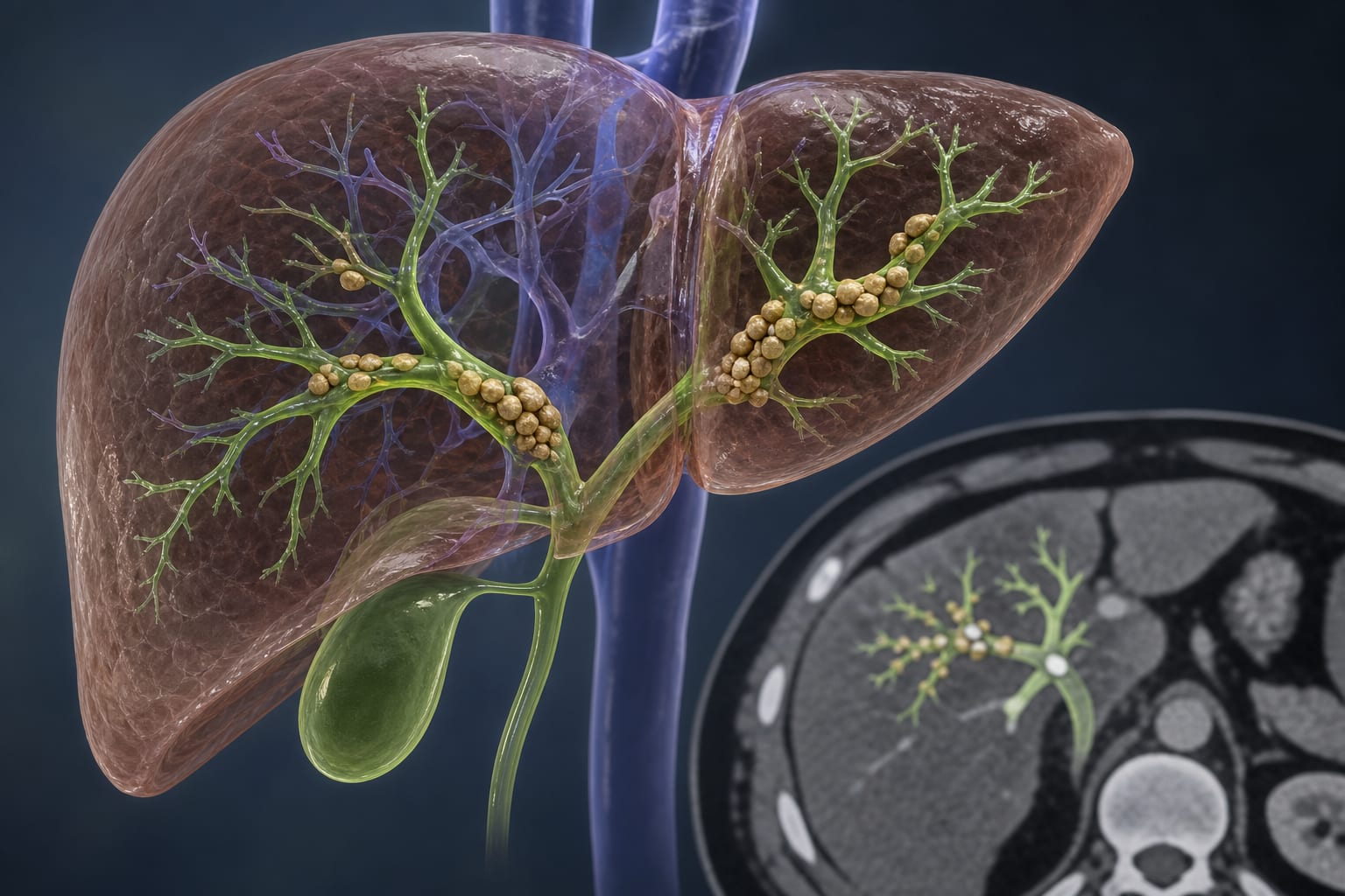
- Hepatolithiasis has a high recurrence rate of postoperative calculi.
- 3D visualization improves the understanding of liver anatomy and intrahepatic duct system.
- One-step PTCSL is less invasive compared to traditional hepatectomy.
- Ultrasound imaging is used to guide the PTCSL procedure.
- The study retrospectively analyzes treatment outcomes using modified rigid choledochoscope lithotripsy.
Guideline-Based Recommendations
Diagnosis
- Diagnosis of hepatolithiasis via preoperative imaging techniques such as US, CT, MRI, MRCP, or PTC.
Management
- Utilization of one-step PTCSL with 3D visualization for treatment.
Monitoring & Follow-up
- Postoperative monitoring for complications and recurrence of stones.
Risks
- Potential for intraoperative injuries and postoperative complications.
Patient & Prescribing Data
Patients with Child–Pugh grade A or B liver function without severe comorbidities.
Modified rigid choledochoscope lithotripsy under 3D visualization improves surgical outcomes.
Clinical Best Practices
- Incorporate 3D visualization in surgical planning for complex hepatolithiasis.
- Ensure thorough preoperative imaging assessment to guide treatment.
- Utilize minimally invasive techniques to reduce recovery time.
Related Resources & Content