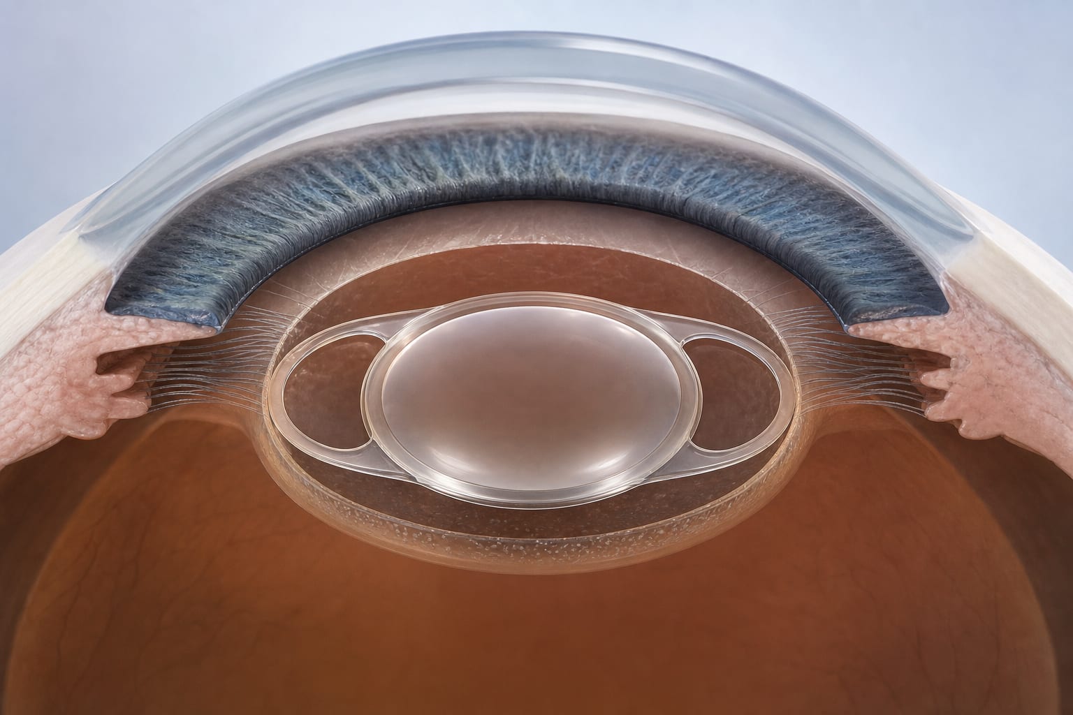
Bag-in-the-lens technique
-
By
-
Shervine Ameli
-
Iva Krolo
-
Silke Oellerich
-
Luc Van Os
-
Marie-José Tassignon
-
Sorcha Ní Dhubhghaill
-
May 8, 2026
Clinical Scorecard: Intraocular Lens Placement Using the Bag-in-the-Lens Method
At a Glance
| Category | Detail |
|---|
| Condition | |
| Key Mechanisms | Prevention of posterior capsular opacification (PCO) through dual capsulorhexis technique, ensuring optimal lens positioning. |
| Target Population | |
| Care Setting | |
Key Highlights
- BIL technique optimizes lens centration and prevents PCO
- Utilizes both anterior and posterior capsulorhexis for lens stabilization, crucial for preventing PCO
- BIL IOL captures residual lens epithelial cells to maintain visual clarity
- Current standard model is the foldable Morcher 89A lens, which enhances stability
- Surgical technique includes calibrated continuous curvilinear capsulorhexes
Guideline-Based Recommendations
Diagnosis
Management
Monitoring & Follow-up
- Regular follow-up for visual acuity and signs of PCO post-surgery, ideally at 1 week, 1 month, and 3 months.
Risks
Patient & Prescribing Data
Patients with cataracts requiring surgical intervention
BIL technique is associated with improved stability and reduced risk of PCO
Clinical Best Practices
- Ensure proper centration of anterior capsulorhexis using a ring caliper
- Utilize dual capsulorhexis to confine lens epithelial cells
- Monitor for postoperative complications and visual outcomes, including specific signs of retinal detachment and increased IOP.
Related Resources & Content