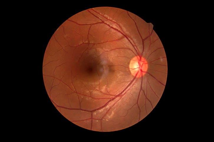
Evaluation of Changes in Optic Nerve Head and Macular Perfusion Before and After COVID-19 Onset
By
Seyfettin Erdem
Sedat Ava
Mehmet Emin Dursun
Mine Karahan
Leyla Hazar
Ugur Keklikci
February 28, 2026
Clinical Scorecard: Evaluation of Changes in Optic Nerve Head and Macular Perfusion Before and After COVID-19 Onset
At a Glance
Category Detail
Condition COVID-19-associated ocular microvascular alterations Key Mechanisms SARS-CoV-2 infection induces endothelial inflammation, dysfunction, and hypercoagulability leading to retinal microvascular changes Target Population Patients who contracted and recovered from COVID-19 Care Setting Ophthalmology clinics with access to optical coherence tomography angiography (OCTA)
Key Highlights
SARS-CoV-2 infects ocular tissues expressing ACE2 receptors and has been detected in retinal biopsies and tear fluid. COVID-19 causes retinal microvascular changes including reduced vessel density in optic nerve head and macular regions. OCTA enables non-invasive, high-resolution longitudinal assessment of retinal microcirculation before and after COVID-19 infection. Guideline-Based Recommendations
Diagnosis
Use polymerase chain reaction (PCR) testing to confirm COVID-19 infection. Perform comprehensive anterior and posterior segment ocular examinations including best-corrected visual acuity. Employ OCTA imaging to quantitatively assess retinal and optic nerve head vessel densities and foveal avascular zone. Management
Exclude patients with confounding ocular or systemic vascular diseases to isolate COVID-19 effects. Monitor recovered COVID-19 patients for retinal microvascular alterations using OCTA. Consider longitudinal OCTA imaging to detect early microvascular changes that may not be clinically evident. Monitoring & Follow-up
Repeat OCTA imaging at least one month post-COVID-19 recovery to evaluate changes in vessel density. Assess vessel density in superficial and deep capillary plexuses, optic nerve head, and radial peripapillary capillary network. Ensure image quality with signal strength >50 and absence of segmentation or motion artifacts. Risks
Potential for retinal vascular occlusions and microvascular damage due to COVID-19-induced hypercoagulability and endothelial dysfunction. Risk of vision-threatening complications secondary to microvascular impairment in optic nerve head and macula. Patient & Prescribing Data
20 patients with baseline OCTA prior to COVID-19 infection who subsequently recovered from the disease
Significant reductions in optic nerve head and radial peripapillary capillary vessel densities were observed post-COVID-19, indicating microvascular impairment despite clinical recovery.
Clinical Best Practices
Obtain baseline OCTA imaging in patients at risk to enable longitudinal comparison post-COVID-19 infection. Exclude confounding ocular and systemic diseases to accurately attribute microvascular changes to COVID-19. Use paired statistical analyses for within-subject longitudinal OCTA data to detect subtle microvascular alterations. Ensure rigorous image quality control with independent specialist review to validate OCTA measurements. Related Resources & Content