Intraoperative functional brain mapping for glioma surgery: a comprehensive review of the University of California San Francisco mapping protocol
By
Jia-Shu Chen
Brandon Bergsneider
Alexander F. Haddad
Ramin A. Morshed
Shawn L. Hervey-Jumper
Jacob S. Young
Mitchel S. Berger
June 13, 2026
Clinical Scorecard: Intraoperative Brain Mapping Techniques for Glioma Resection: An In-Depth Analysis of the UCSF Protocol
At a Glance
Category Detail
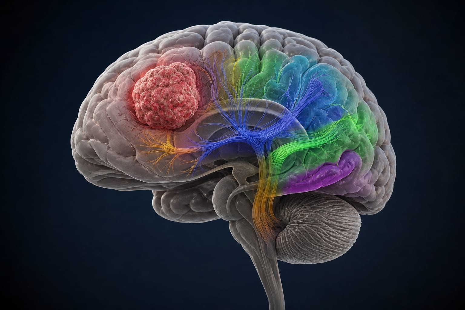
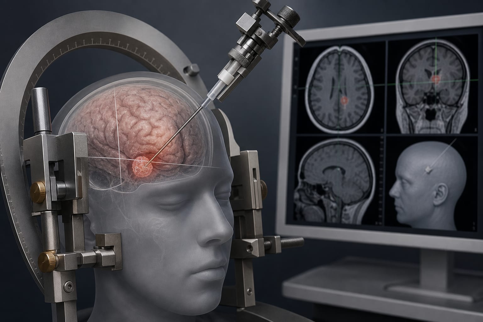
Condition Glioma Key Mechanisms Intraoperative functional brain mapping to identify motor and language pathways. Target Population Patients with newly-diagnosed adult-type diffuse glioma, particularly those with tumors involving motor and/or language areas. Care Setting Neurosurgical operating room.
Key Highlights
Maximal resection of contrast-enhancing and FLAIR borders is the gold standard for glioma treatment. Intraoperative mapping is associated with fewer neurological deficits and higher rates of maximal resection. Awake and asleep mapping techniques are utilized to enhance patient safety and comfort. Preoperative imaging with DTI and MEG is critical for surgical planning. The onco-functional outcome (OFO) classification scheme helps assess resection outcomes. Guideline-Based Recommendations
Diagnosis
Utilize MRI with and without gadolinium, DTI, and MEG for preoperative assessment. Management
Perform maximal resection while preserving functional status. Monitoring & Follow-up
Assess neurological function postoperatively to evaluate for deficits. Risks
Increased risk of neurological deficits with larger resections, especially near functional cortex. Patient & Prescribing Data
Adults with diffuse gliomas, particularly those with IDH mutant astrocytomas and oligodendrogliomas.
Supratotal resection beyond FLAIR margins is associated with lower recurrence risk and improved survival.
Clinical Best Practices
Implement intraoperative motor and language mapping to minimize neurological deficits. Utilize asleep conditions for motor mapping to enhance accuracy and patient comfort. Incorporate DTI and MEG in preoperative planning to improve surgical outcomes. Related Resources & Content