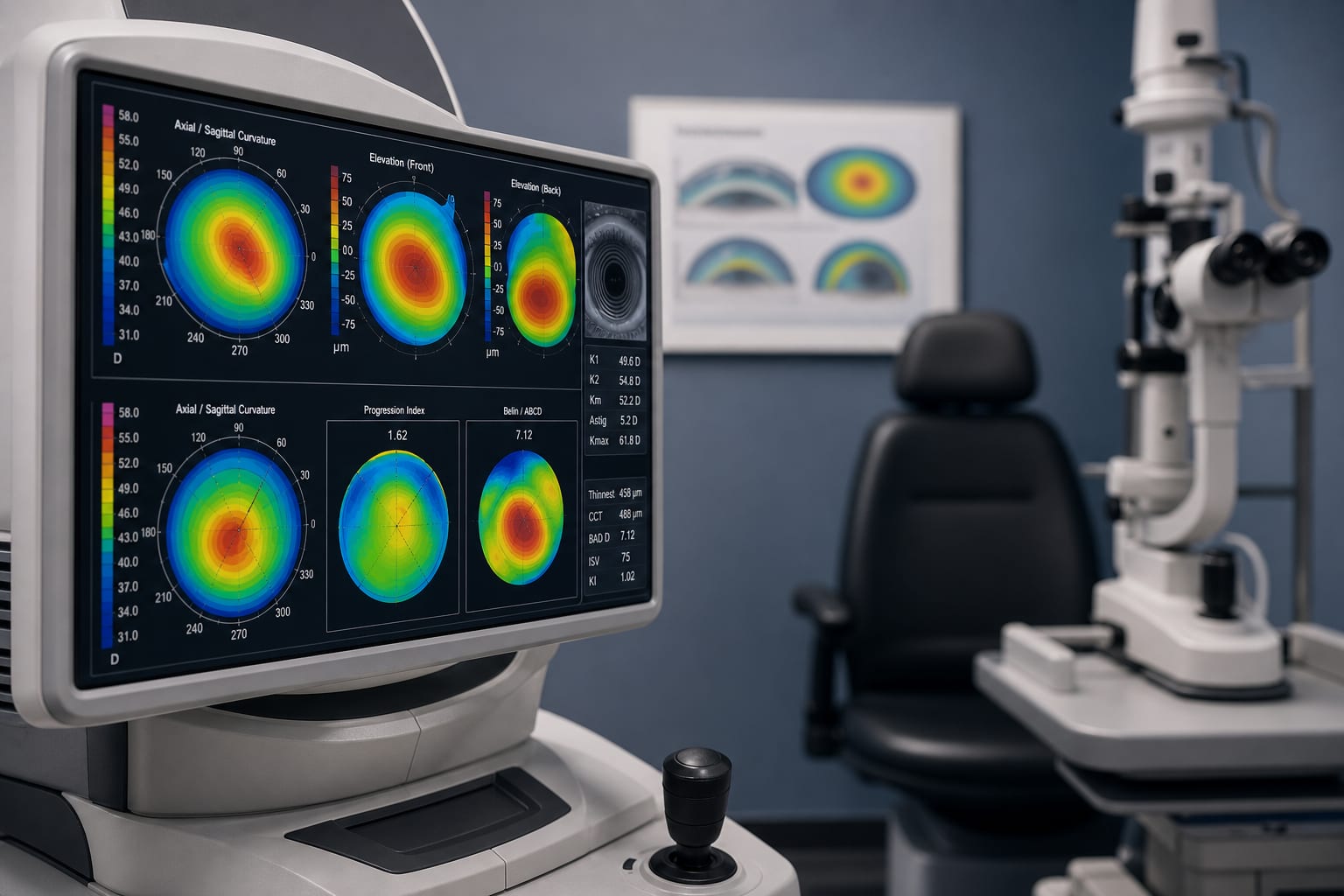
Evaluation of Corneal Thickness Using the New Revo FC Optical Coherence Tomographer Compared to the Pentacam Scheimpflug Imaging System - Scorecard - MDSpire
Clinical Scorecard: Evaluation of Corneal Thickness Using the New Revo FC Optical Coherence Tomographer Compared to the Pentacam Scheimpflug Imaging System
At a Glance
Category
Detail
Condition
Corneal thickness measurement
Key Mechanisms
Assessment of corneal thickness for diagnosis and management of ophthalmic conditions
Target Population
Patients with healthy eyes, excluding those with ocular diseases or previous surgeries
Care Setting
Ophthalmology clinics
Key Highlights
Corneal thickness is crucial for diagnosing conditions like keratoconus and planning refractive surgeries.
The Revo FC OCT offers improved speed and measurement reliability compared to the Pentacam system.
Mean central corneal thickness (CCT) was 545.52 μm with Pentacam and 539.36 μm with Revo FC OCT.
Both systems showed a high correlation in CCT measurements (correlation coefficient 0.99).
Study included 74 patients with a mean age of 36.50 years.
Guideline-Based Recommendations
Diagnosis
Use corneal thickness measurements to evaluate keratoconus and other corneal pathologies.
Management
Ensure adequate corneal thickness before refractive surgery to minimize risks.
Monitoring & Follow-up
Regularly monitor corneal thickness in patients with known corneal conditions.
Risks
Inadequate corneal thickness can lead to postoperative complications such as iatrogenic keratectasia.
Patient & Prescribing Data
Patients with healthy eyes, aged 18-65 years, without ocular diseases or previous surgeries.
Accurate corneal thickness measurements are essential for safe refractive surgery planning.
Clinical Best Practices
Calibrate devices according to manufacturer guidelines before measurements.
Perform thorough ophthalmological examinations prior to corneal thickness assessment.
Use pachymetric maps for detailed analysis of corneal biomechanics.