Analysis of ultrasonic characteristics in 12 cases of ovarian Sertoli–Leydig cell tumour
-
By
-
Liyang Shao
-
Weihong Dong
-
Shuang Li
-
Qiongrui Zhao
-
Tingting Liu
-
Kaikai Li
-
Tian Wu
-
Yu Wang
-
Haohui Zhu
-
Ruili Wang
-
June 23, 2026
-
Clinical Scorecard: Ultrasonic Imaging Features in Twelve Cases of Ovarian Sertoli–Leydig Cell Tumors
At a Glance
| Category | Detail |
|---|
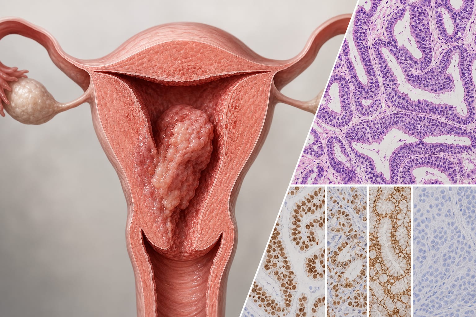
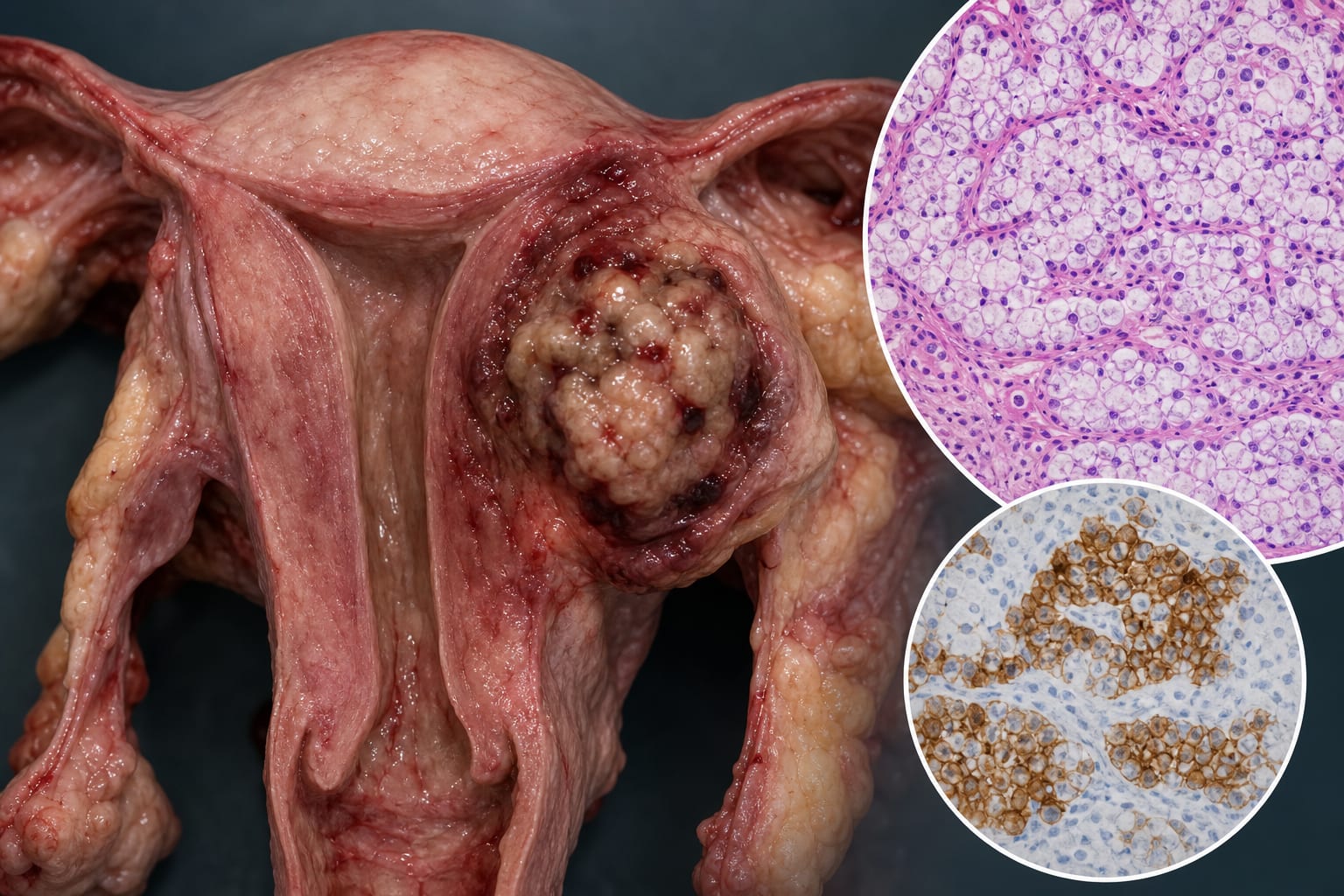
| Condition | Ovarian Sertoli–Leydig Cell Tumors |
| Key Mechanisms | Tumor cells produce androgens leading to hyperandrogenaemia and associated clinical features. |
| Target Population | Younger females, median age approximately 30 years. |
| Care Setting | Ultrasound imaging for ovarian neoplasms. |
Key Highlights
- SLCTs predominantly present as unilateral, well-defined solid or cystic-solid adnexal masses.
- Notable peripheral vascularity and surrounding ball Doppler pattern observed in many cases.
- Elevated preoperative testosterone levels noted in all patients with accessible data.
Guideline-Based Recommendations
Diagnosis
- Ultrasound is the principal screening modality for ovarian neoplasms.
Management
- Surgical intervention is necessary for definitive diagnosis and treatment.
Monitoring & Follow-up
- Regular follow-up and monitoring of hormone levels may be warranted.
Risks
- Malignant potential of SLCTs necessitates prompt diagnosis and intervention.
Patient & Prescribing Data
12 individuals diagnosed with ovarian SLCTs.
Histopathological confirmation is essential for diagnosis.
Clinical Best Practices
- Consider SLCTs in younger patients with hyperandrogenic features.
- Utilize ultrasound to assess tumor morphology and vascularity.
Related Resources & Content